Abstract
Prostatic artery (PA) origination from a common trunk with the superior vesical artery (SVA) is a frequent cause of technical difficulties in PA catheterization for PA embolization (PAE). These difficulties, which substantially increase the operative time, radiation dose, cost, and technical failure rate of PAE, can often be overcome by the utilization of a steerable microcatheter (MC) with a tip that can be manually adjusted at an angle that optimally conforms to the shape and origin of the common vesicoprostatic trunk. Adjunctive techniques that can be applied when the steerable MC fails to engage the PA include: 1) the protective temporary embolization of the SVA so that a permanent embolic can be redirected into the PA; 2) PAE via collaterals between superior vesical branches and the PA; and 3) embolization from a proximal position of the MC near the PA orifice to exploit preferential flow to the PA. In the authors’ recent experience, the utilization of a steerable MC with and without adjunctive techniques (in 12 and 23 patients, respectively) resulted in a 35% increase in the technically successful embolization of PAs originating from vesicoprostatic trunks with no significant complications. Familiarization with alternative devices and techniques may substantially improve the technical outcome of PAE in cases with challenging arterial anatomy.
Keywords: Angiography, microcatheter, prostatic artery, prostatic artery embolization, vesicoprostatic trunk
Main points
• Prostatic artery (PA) origination from the anterior division of the internal iliac artery in the form of a common trunk with the superior vesical artery (i.e., a vesicoprostatic trunk) is frequently encountered.
• This vesicoprostatic trunk may be clinically relevant to PA embolization because of the frequently associated difficulties arising from it during the procedure.
• These difficulties may result in increased operative times, radiation doses, and costs and even in technical failure of the procedure.
• A steerable microcatheter with a tip that can be manually adjusted to an angle of 0°–180° can be employed with or without adjunctive techniques to overcome the aforementioned difficulties.
Prostatic artery (PA) origination from a common vesicoprostatic trunk with the superior vesical artery (SVA) (referred to as type 1 PA origination, according to a widely used angiographic classification) is the most prevalent (or second most, depending on the population in question) variety of PA origination., Compared with other types of PA origination, type 1 is associated with significantly more technical difficulties during attempted PA catheterization for PA embolization (PAE). These difficulties, mainly caused by a short and cranially oriented vesicoprostatic trunk, with or without an unfavorable angle of origin of the PA from this common trunk, may lead to prolonged operative times, increased radiation doses for staff and patients, and the need for additional microcatheters (MCs) and microguidewires (MGWs)., The rate of unsuccessful attempts at superselective catheterization of the PA is also significantly higher for type 1 origination compared with all other types combined, and this eventually translates into unilateral (rather than bilateral) PAE procedures with suboptimal clinical outcomes.
Among other approaches, the utilization of a steerable MC with a tip manually adjustable by the operator to angles of 0°–180°, has been briefly reported in the literature,, as an option to address the challenges associated with vesicoprostatic trunks during PAE. There is likely room for refinement and the application of adjunctive techniques in the deployment of the steerable MC to further improve the technical outcomes of PAEs. The aim of this work is therefore to describe the utilization of a steerable MC and of adjunctive techniques for PAE in the context of anatomically challenging vesicoprostatic trunks. The clinical efficacy and safety of the described techniques is also briefly evaluated.
Methods
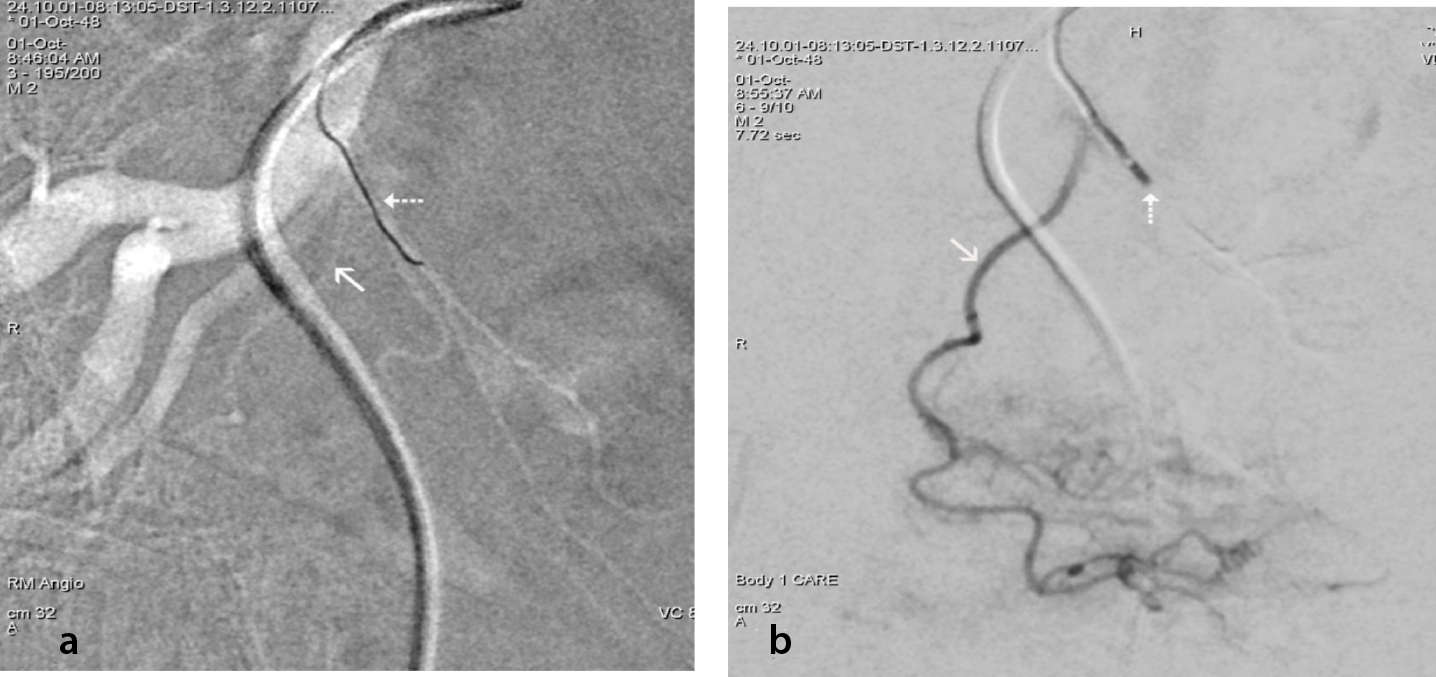
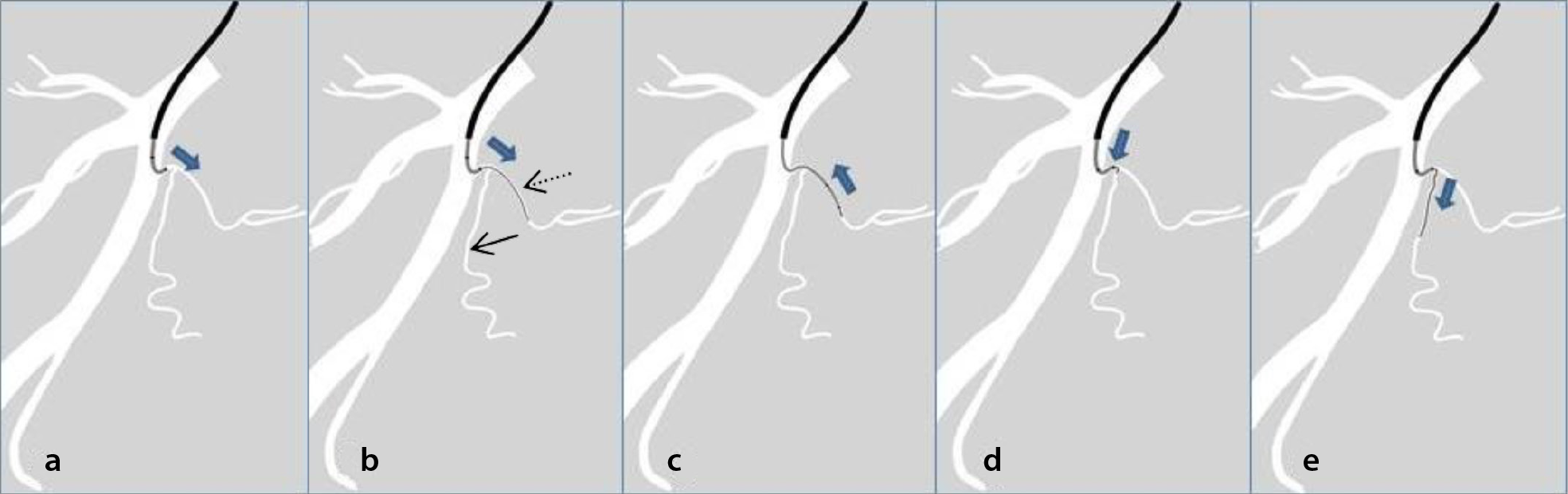
In the centers of the authors, a steerable 2.4 French (Fr) MC (SwiftNINJA, Merit Medical Systems, Inc., South Jordan, UT, USA) was deployed when catheterization of the common vesicoprostatic trunk proved impossible after attempts of approximately 3 min duration with the initial standard combination of devices. This combination included a 2 Fr MC (Parkway Soft-Asahi Intecc Co., Japan) and a double-angled MGW (0.016” Meister, Asahi Intecc Co.). Compared with the standard device, the tip of the steerable MC can not only be adjusted so that it is optimally oriented to the orifice of the common vesicoprostatic trunk but also provides better support and a steadier angle that is not prone to straightening upon insertion of the MGW. The tip of the steerable MC was locked at the desired angle and catheterization was attempted. After initial engagement of the orifice of the common vesicoprostatic trunk with the MGW, the MGW tended to advance more easily into the SVA than into the PA. After distal advancement of the MGW into the SVA, the angled tip of the steerable MC was “unlocked,” and the device was advanced distally over the MGW and into the SVA to secure this first step of catheterization. Subsequently, the MC–MGW combination was slowly retracted under fluoroscopy until it reached the origin of the PA. Catheterization of the latter was then attempted with appropriate rotation of the double-angled MGW and, as necessary, with a new adjustment of the tip of the MC so that it was angled and “locked” in the direction of PA origination (, ). The same double-angled MGW used in the initial attempts was combined with the steerable MC. Occasionally, however, if appropriately angled, the steerable MC alone (without the MGW) could be advanced to engage first the common vesicoprostatic trunk and then the PA (, ). A high-quality “roadmap” image greatly facilitated the identification of the origins of the target arteries.
When attempts with a steerable MC and MGW proved fruitless, the following techniques were applied:
1. Protective temporary embolization of the SVA. This can be applied when the MC easily advances into the SVA but cannot be redirected into the PA. In the current work, a temporary embolic agent (EmboCube Gelatin, Merit Medical) with particles of a hydrated size of 2.5 mm was injected through the MC into the SVA. Care was taken to avoid a too-distal or too-proximal occlusion, including at the PA origin. After angiographic confirmation of an SVA occlusion, the MC was withdrawn just proximal to the PA origin. A permanent embolic (composed of microspheres) was then slowly injected, and its redirection into the PA was fluoroscopically documented (). Care is required to avoid backflow into the anterior division of the internal iliac artery. Occasionally, resistance to the advancement of the MGW in the SVA after its occlusion forces the MGW to enter an otherwise non-selectable PA. At this point, superselective catheterization of the PA can be accomplished. Finally, although gelatin is considered a temporary embolic, the authors confirm the patency of the contralateral SVA prior to the protective occlusion of the ipsilateral SVA.
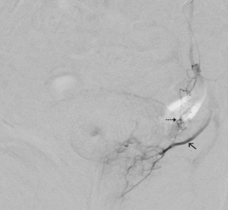
2. PAE via collaterals. Descending SVA branches with rich anastomoses to the PA may occasionally be encountered. Owing to their usually obtuse angle of origin from the SVA, superselective catheterization and the distal advancement of the MC into these branches is much easier than catheterization of the PA. If a slow manual contrast injection in these branches reveals substantial prostatic opacification and negligible perfusion of the bladder wall, PAE can be safely performed through these descending SVA branches ().
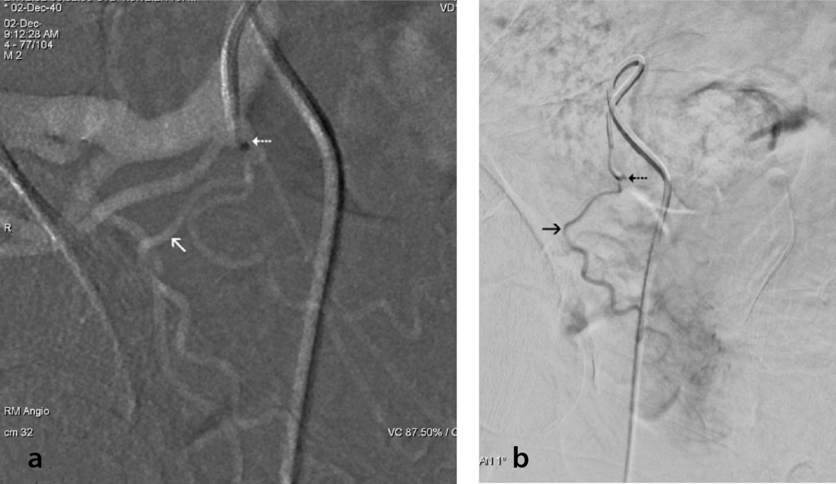
3. Embolization from a proximal position of the MC near the PA orifice that exploits preferential flow to the PA. This can be applied when all previous options have failed and when the tip of the steerable MC can only reach the orifice of the PA without engaging it. In the current work, with appropriate rotation of the MC and/or adjustments of the angle of its tip, the latter was positioned against the PA orifice. Angiograms were acquired during slow manual contrast injections to ensure that only the PA was opacified (). Embolization was then slowly performed and stopped when a sub-stasis of flow was observed in the PA.
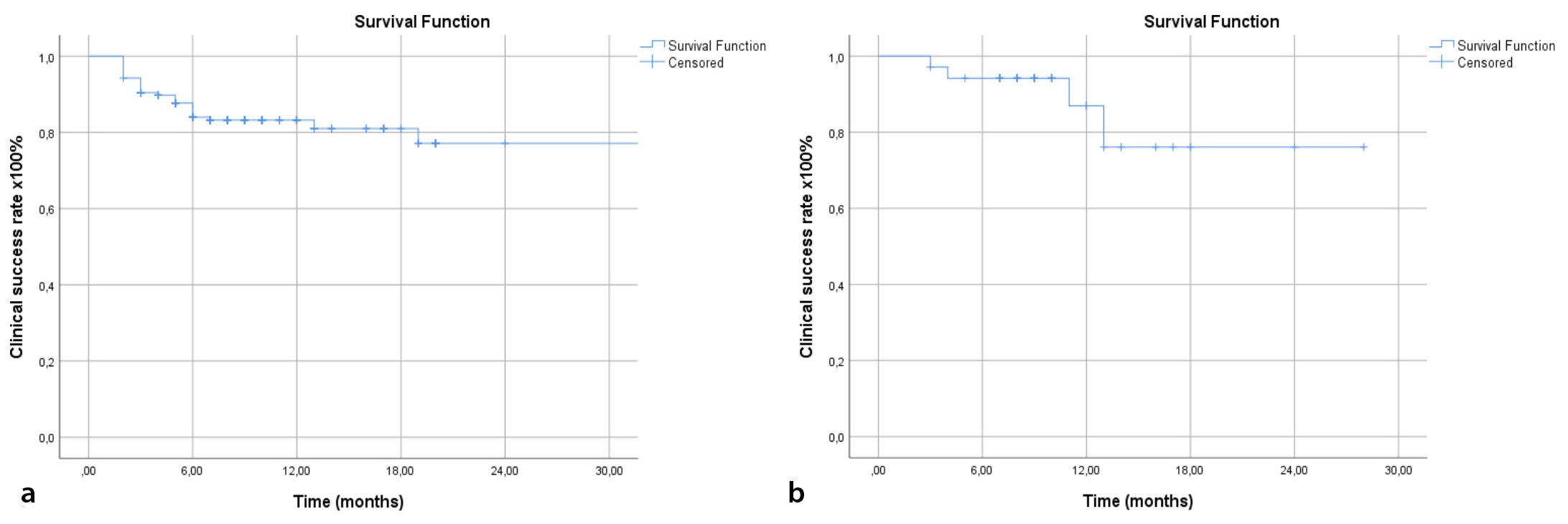
Of a total of 157 patients with benign prostatic hyperplasia (314 pelvic sides) treated with PAE in the centers of the authors during the last 3 years (), a common vesicoprostatic trunk was observed in 101 pelvic sides (32.2%). In cases with a double or triple PA per pelvic side, only the most prominent PA was registered. All patients were informed in detail of both the standard and the adjunctive PAE techniques and provided written informed consent prior to the procedure. Of the 101 cases with vesicoprostatic trunks, PAE was accomplished with a standard MC in 59 cases, PA catheterization required additional utilization of steerable MC with no adjunctive technique in 23 cases, and a steerable MC and adjunctive techniques were eventually employed in 12 cases (). In the remaining 7 cases, utilization of one of the aforementioned approaches either failed or adjunctive techniques were contraindicated, and the patients underwent unilateral PAE. All 35 patients who were successfully treated with a steerable MC, with or without adjunctive techniques, underwent bilateral PAE, and the clinical success rate 1 year post-PAE was 87%. No major complications were observed. Minor complications were observed in 6 of the 35 patients. The technical success rate within the entire cohort of 157 patients was 96.8% (bilateral PAE in 128 patients, unilateral PAE in 24 patients, and technical failure/no PAE in 5 patients). The clinical success rate 1 year post-PAE was 83.7%, and complications (minor only) were encountered in 25 of the 157 patients (, ). Finally, imaging from and the clinical outcomes of the 35 patients who were treated with the steerable MC, with or without adjunctive techniques, were comparable with a previous series from the same centers within which only conventional catheterization techniques were applied.
Table 1. Demographic and clinical features of the patients
Variable | Value for all patients with PAE treated in the two centers (n = 157) | Value for the subgroup treated with steerable MC ± adjunctive techniques (n = 35) |
|---|---|---|
Age (y; mean ± SD) | 71.2 ± 10.3 | 73.1 ± 11.2 |
BMI (mean ± SD) | 26.6 ± 2.6 | 27.1 ± 3.1 |
PV (mL; mean ± SD) | 87.1 ± 48.2 | 77.4 ± 41.2 |
Indication for PAE (proportion of patients) | ||
Moderate LUTS | 54/157 | 11/35 |
Severe LUTS | 57/157 | 14/35 |
Indwelling bladder catheter | 40/157 | 9/35 |
Hemorrhage of prostatic origin | 6/157 | 1/35 |
PAE, prostatic artery embolization; MC, microcatheter; SD, standard deviation; BMI, body mass index; PV, prostate volume; LUTS, lower urinary tract symptoms; y, years.
Table 2. Additional data about the adjunctive techniques for PAE
Adjunctive technique | Number of patients | Embolic material | Percentage of prostatic infarction of the treated lobe* | Clinical success 1 year post-PAE (proportion of patients) |
Protective SVA embolization | 6 | Embosphere (100–300 μm) | 5%–55% | 6/6 |
PA embolization via collaterals | 3 | Embosphere (100–300 μm) | 31%–39% | 3/3 |
Proximal PA embolization and exploitation of preferential flow to the PA | 3 | Embosphere (300–500 μm) | 0%–26% | 2/3 |
*Percentage of prostatic infarction = the volume of infarcts in the treated lobe/the total volume of the treated lobe (infarcts were evaluated with contrast-enhanced ultrasound). PAE, prostatic artery embolization; SVA, superior vesical artery; PA, prostatic artery.
Discussion
According to a practical and widely accepted approach, PA origination can be angiographically classified into five types. In type 1, the PA and the SVA share a common origin (trunk) from the anterior division of the internal iliac artery. In type 2, the PA originates from the anterior division of the internal iliac artery (separately from the SVA). Type 3 describes PA origination from the obturator artery, while type 4 indicates origination from the internal pudendal artery. Finally, type 5 includes rare PA origins, such as origination from the accessory pudendal or aberrant obturator artery. Among other vasculo-anatomical factors, PA origination type affects the technical outcome of PAE, with type 1 most often associated with difficult or failed PA catheterizations.
Compared with standard MCs, the utilization of a steerable MC appears to significantly increase the chances of the successful catheterization of type 1 PA originations—particularly in cases of short, cranially oriented vesicoprostatic trunks with an acute angle of PA origination,,,—by up to approximately 35%, according to the experience presented herein. The steerable MC can also address additional challenges that often coexist with type I PA origin, such as a tortuous and ectatic anterior division of the internal iliac artery or a base catheter facing posterolaterally instead of anteromedially. When preinterventional computed tomographic angiography reveals such a challenging anatomy, it may be more practical to begin with a steerable rather than a standard MC; however, the financial aspects of this approach should be further investigated.
With the adjunctive techniques described herein, PAE can be performed even when superselective catheterization of the PA is impossible. The following technique-specific comments can also be made: 1) protective coiling of the SVA has been described before and, despite its permanent nature, is considered a safe technique. However, since protective occlusion of the SVA is only needed during the few minutes of the injection of microspheres into the PA, the authors prefer a temporary embolic agent for the protection of the SVA, with the potential for complete SVA recanalization in the following days or weeks. Moreover, uniformly cut gelatin particles with a standardized hydrated size probably ensure a more controlled and precise occlusion compared with the gelfoam slurry prepared by the operator; 2) collaterals between vesical and prostatic arteries are not uncommon, but they can only rarely serve as pathways for safe and effective PAE. Distal advancement of the MC in the collaterals and slow, controlled manual contrast injections are required to confirm abundant flow to the prostate and the absence of bladder wall opacification; and, finally 3) it should be acknowledged that the exploitation of preferential flow to the PA is a suboptimal PAE technique that should be applied only when previous options have failed. Relatively larger microspheres with a diameter of 300–500 microns (rather than 100–300 microns) are preferred for this last option to minimize the risk of ischemic complications in the case of reflux to the SVA or to the more distal branches of the anterior division of the internal iliac artery.
Other options to address the aforementioned difficulties are either not widely available—such as utilization of a robotic c atheter—or are more complex and invasive—such as the combination of a larger sheath and a “buddy wire”. The adjunctive techniques presented herein appear to be simpler, more widely available, and affordable, as they can be applied not only with steerable but also with standard MCs.
In conclusion, familiarization with the application of a steerable MC and with the adjunctive techniques described herein may substantially improve the technical outcome of PAE in cases of anatomically challenging vesicoprostatic trunks.
Footnotes
Acknowledgements
The authors are grateful to Mr. Konstantinos Chouchos, MSc, and Mrs Maria-Theodora Moschouri (medical student) for their valuable assistance in the preparation of this manuscript.
Conflict of interest disclosure
The authors declared no conflicts of interest.
References
de Assis AM, Moreira AM, de Paula Rodrigues VC, et al. Pelvic arterial anatomy relevant to prostatic artery embolisation and proposal for angiographic classification.Cardiovasc Intervent Radiol. 2015;38(4):855-861.
Moschouris H, Stamatiou K, Tzamarias S, et al. Angiographic imaging of prostatic artery origin in a greek population and correlation with technical and clinical aspects of prostatic artery embolization.Cureus. 2023;15(9):e45941.
Boeken T, Gautier A, Moussa N, et al. Impact of anatomy type of prostatic artery on the number of catheters needed for prostatic artery embolization.Diagn Interv Imaging. 2021;102(3):147-152.
Hoffmann JC, Minkin J, Primiano N, Yun J, Eweka A. Use of a steerable microcatheter during superselective angiography: impact on radiation exposure and procedural efficiency.CVIR Endovasc. 2019;2(1):35.
Mouli S, Hohlastos E, Salem R. Prostate artery embolization.Semin Intervent Radiol. 2019;36(2):142-148.
Moschouris H, Stamatiou K, Malagari K, et al. The value of contrast-enhanced ultrasonography in detection of prostatic infarction after prostatic artery embolization for the treatment of symptomatic benign prostatic hyperplasia.Diagn Interv Radiol. 2019;25(2):134-143.
Bagla S, Isaacson AJ. Tips and tricks for difficult prostatic artery embolization.Semin Intervent Radiol. 2016;33(3):236-239.
Richardson AJ, Acharya V, Kably I, Bhatia S. Prostatic artery embolization: Variant origins and collaterals.Tech Vasc Interv Radiol. 2020;23(3):100690.
Supplementary Materials
Supplementary Video 1. Despite suboptimal support from and unfavorable rotation of the base catheter, the steerable MC can be appropriately angled and directed into the vesicoprostatic trunk without a MGW. MC, microcatheter, MGW, microguidewire.
DownloadSupplementary Table 1. Complications encountered in the patients of this work
Complication* | Number of patients (%) | |
|---|---|---|
- | For the entire patient cohort (n = 157) | For the subgroup treated with steerable MC ± adjunctive techniques (n = 35) |
Haematospermia | 3 (1.9) | - |
Haematuria | 2 (1.3) | 1 (2.8) |
Penile ulcers (small, ischemic) | 2 (1.3) | - |
Acute urinary retention | 9 (5.7) | 4 (11.5) |
Prostatic tissue expulsion | 1 (0.6) | - |
Rectal bleeding | 2 (1.3) | - |
Urinary tract infection | 2 (1.3) | - |
Inguinal haematoma | 4 (2.5) | 1(2.8) |
Total | 25 (15.9) | 6 (17.1) |
*All complications were considered minor, because they required no hospitalization and were self-limiting, or resolved with conservative treatment. MC, microcatheter.
Supplementary Figure 1. Schematic drawing showing usual maneuvers for prostatic artery (PA) catheterization in challenging case of vesicoprostatic (VP) trunk. (a) The tip of the steerable microcatheter (MC) is manually angulated towards the orifice of the VP trunk and locked. (b) The microguidewire (MGW) is inserted; more often than not, it advances into the superior vesical artery (black dotted arrow) instead of the PA (black arrow). (c) The tip of the MC is unlocked and the latter is inserted into the superior vesical artery over the MGW. (d) The MC and MGW are slowly withdrawn close to the origin of the PA; the latter is catheterized with appropriate rotation of the MGW, and, if required, additional angulation and locking of the MC. (e) The MGW is advanced into the PA. All drawings correspond to ipsilateral oblique angiographic projections. Blue arrow indicates the direction of advancement (or withdrawal) of MC and MGW.
Supplementary Video 2. Despite unfavorable rotation of the base catheter, appropriate angulation of the steerable MC can help the operator to direct the MGW into the vesicoprostatic trunk and then into the PA. MC, microcatheter, MGW, microguidewire, PA, prostatic artery.
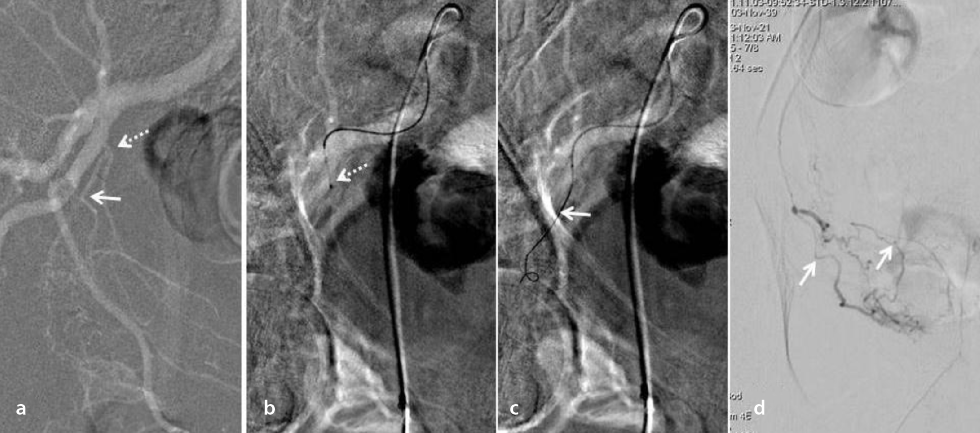
DownloadSupplementary Figure 2. Representative case of utilization of steerable microcatheter (MC) in the context of challenging vesicoprostatic (VP) trunk. Ipsilateral oblique roadmap image (a), shows a short VP trunk (dotted arrow) which originates at 90° angle from the anterior division of the internal iliac artery. The angle of prostatic artery (PA, arrow) origin from the VP trunk is also unfavorable (less than 90°). Similar projections during attempt of catheterization with steerable MC (b, c), show the tip of the MC (dotted arrow, b) angulated towards the orifice of the VP trunk. Despite suboptimal support from the base catheter, the microguidewire (arrow, c) can be directed into the PA. Anteroposterior angiogram (d) after distal advancement of the MC into the PA shows distal prostatic branches (arrows).
Supplementary Figure 3. Kaplan-Meier curves showing the clinical success rates for the entire patient cohort (n = 157) who unterwent PAE in the 2 centers of the authors during the last 3 years (a), and for the subgroup of patients (n = 35) who underwent PAE with steerable MC ± adjunctive techniques (b). PAE, prostatic artery embolization; MC, microcatheter.