
Abstract
Fluorodeoxyglucose (FDG) positron emission tomography (PET)/magnetic resonance imaging (MRI) is an advanced hybrid imaging modality that holds substantial promise in oncologic imaging. FDG PET, a well-established molecular imaging technique, enables assessment of tumor glucose metabolism and is widely utilized for diagnosing and monitoring a range of malignancies; MRI offers superior soft tissue contrast, facilitating precise anatomical localization of PET findings. This pictorial essay presents a series of physiological FDG uptake variants observed in the uterus, fallopian tubes, ovaries, colon, internal anal sphincter, pelvic musculature, and bone marrow. Additionally, it highlights benign conditions such as uterine tumors, vaginal condyloma acuminatum, and infected Bartholin cysts that may exhibit FDG uptake and potentially mimic malignancy. The essay also illustrates FDG-avid post-treatment changes and various infectious and inflammatory lesions such as osteitis pubis, perianal fistulas, and pelvic abscesses that may complicate oncologic interpretation.
Keywords: Benign, bone marrow, bowel, fallopian tube, false positive results, inflammation, magnetic resonance imaging, ovary, positron emission tomography, radiation therapy, uterus
Main points
• Physiological fluorodeoxyglucose (FDG) uptake is frequently observed, particularly in the uterine cavity, fallopian tubes, corpus luteum, colon, and the distal internal anal sphincter.
• Benign processes and post-treatment changes with FDG uptake can result in false-positive interpretations during positron emission tomography/magnetic resonance imaging for oncologic evaluation.
• Increased FDG uptake can be seen in conditions such as perianal fistulas, vaginal condyloma acuminatum, infected Bartholin cysts, osteitis pubis, and inflammatory bowel disease.
• Diffuse or asymmetrical FDG uptake may be present in pelvic muscles following insulin administration or strenuous physical activity.
• Physiological FDG accumulation in the sigmoid colon and rectum varies among individuals and may be increased in patients on metformin therapy, potentially complicating colorectal cancer assessment.
Positron emission tomography (PET) combined with magnetic resonance imaging (MRI) using fluorodeoxyglucose (FDG) is a promising hybrid imaging tool for evaluating gynecologic and genitourinary malignancies. It has a potential pivotal role in staging, treatment planning, recurrence detection, and prognosis assessment. However, interpreting pelvic PET/MRI is often challenging due to physiological uptake patterns and benign mimickers that may resemble pathology. In this pictorial essay, we aim to illustrate incidental FDG uptake in various pelvic structures encountered during routine oncologic PET/MRI examinations.
Normal Physiological Variants
Physiological uptake of FDG in the uterus, fallopian tubes, and ovaries is a common finding on FDG PET/computed tomography (CT), particularly in premenopausal women, and may mimic pathological processes if not carefully interpreted.
Physiological endometrial uptake
FDG uptake in the uterus varies with hormonal changes during the menstrual cycle, especially in premenopausal women. Increased endometrial FDG activity is commonly observed during the menstrual () and ovulatory phases and is considered physiological. This uptake likely reflects the peristaltic activity of the subendometrial myometrium, which facilitates menstrual blood expulsion.
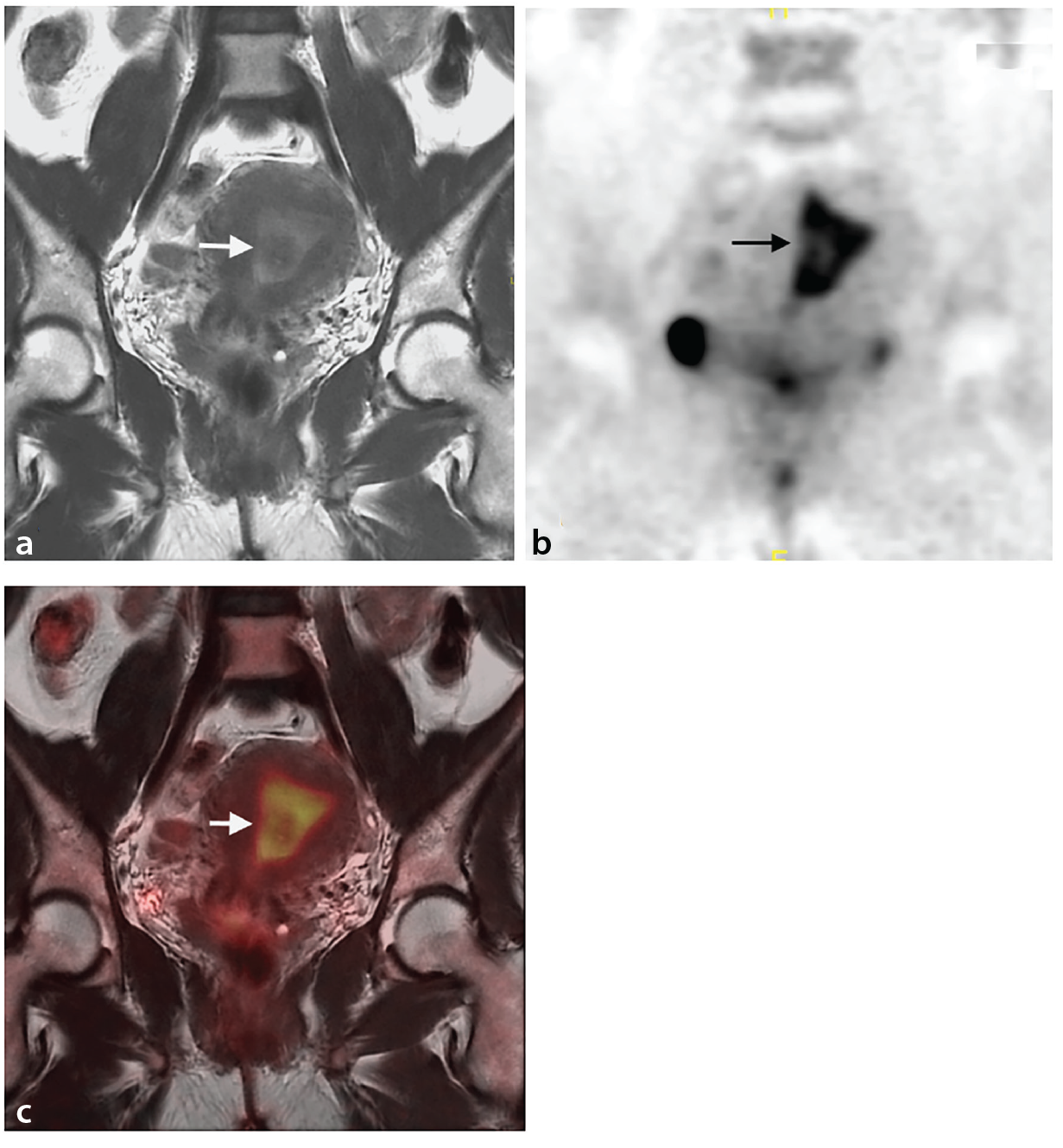
In postmenopausal women, endometrial FDG uptake is typically low. Any focal or intense uptake in this age group should prompt further investigation.
Physiological uptake in fallopian tubes
Studies performed with PET/CT demonstrate that the fallopian tubes may exhibit physiological FDG uptake, particularly in premenopausal women. In a study by Yun et al., bilateral tubal FDG activity was observed in 8.8% of women aged <53 years, most commonly during the mid-menstrual cycle. The uptake appears as tubular, comma-, or tadpole-shaped foci adjacent to the ovaries, corresponding to fallopian tube anatomy.
MRI coregistration and analysis of morphologic features and symmetry can aid differentiation (). In the absence of corresponding structural abnormalities or clinical suspicion, further investigations are usually unnecessary. Misinterpretation of tubal uptake as peritoneal implants, enlarged lymph nodes, or adnexal malignancy may lead to unwarranted procedures.
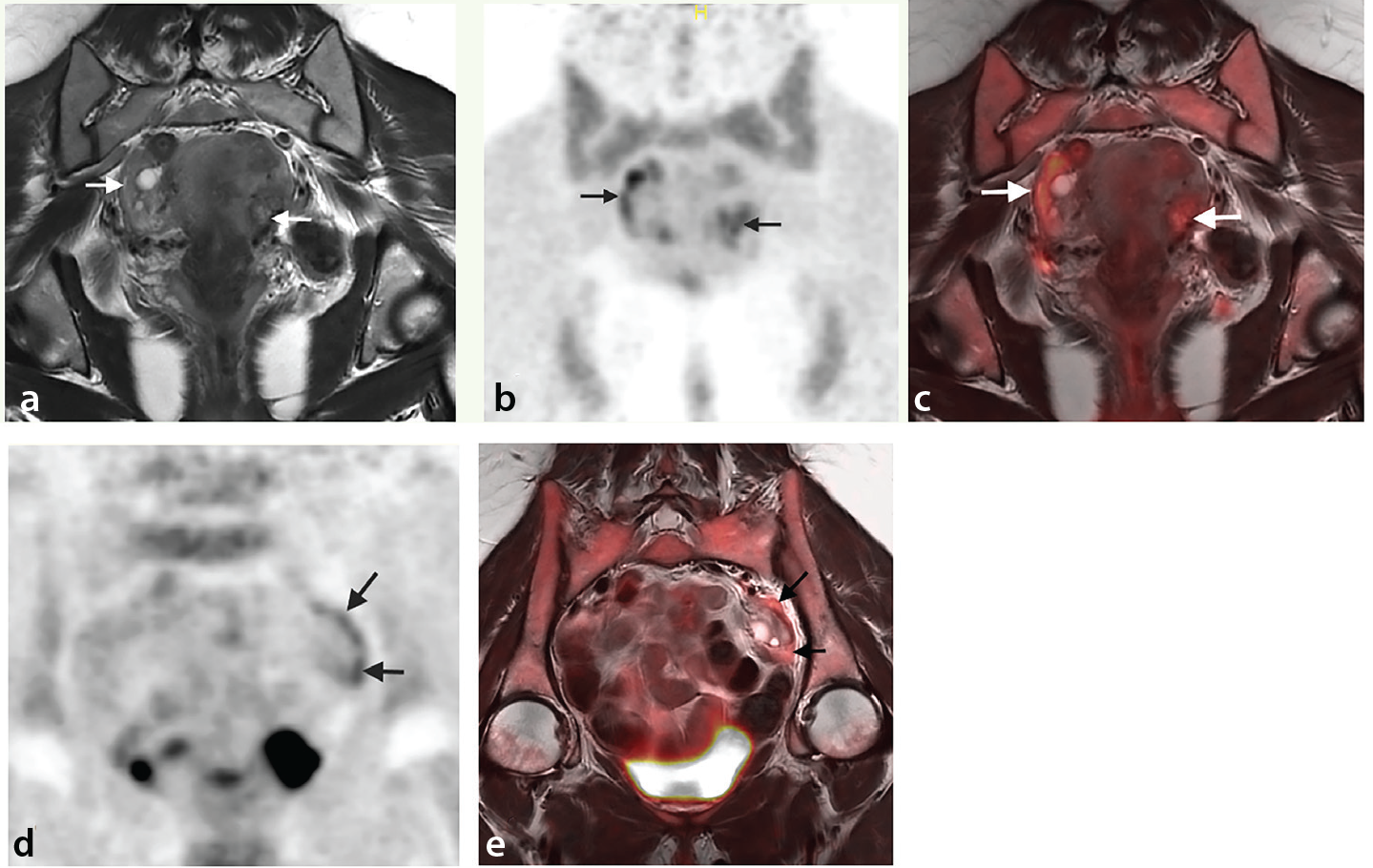
Physiological ovarian uptake
Ovarian FDG uptake fluctuates with the menstrual cycle. A common physiological variant is FDG accumulation in the corpus luteum during the luteal phase. The corpus luteum, a transient endocrine structure formed post-ovulation, produces progesterone and may demonstrate moderate FDG avidity., ,,
This uptake typically appears as a unilateral, focal, rounded lesion (). Although it may mimic malignancy, it is transient and usually resolves in subsequent cycle phases. Correlation with MRI can clarify the nature of the lesion, as corpus luteum cysts appear as unilocular structures (1–3 cm) with thick, crenulated enhancing walls. Presence of blood products may cause signal heterogeneity in the corpus luteum., ,,
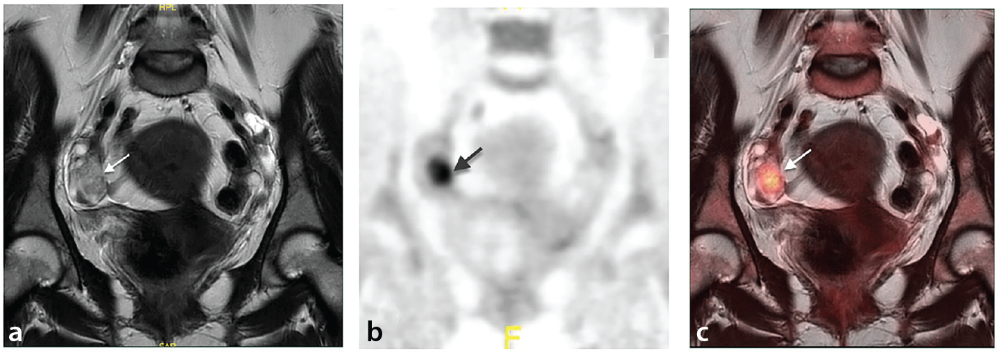
Physiological bowel activity on fluorodeoxyglucose positron emission tomography/magnetic resonance imaging
FDG accumulation is frequently observed in the colon and rectum, typically in a diffuse, linear pattern (). However, focal or segmental uptake may also be present. Rectal FDG activity can pose diagnostic challenges, and correlation with clinical data and MRI is essential.
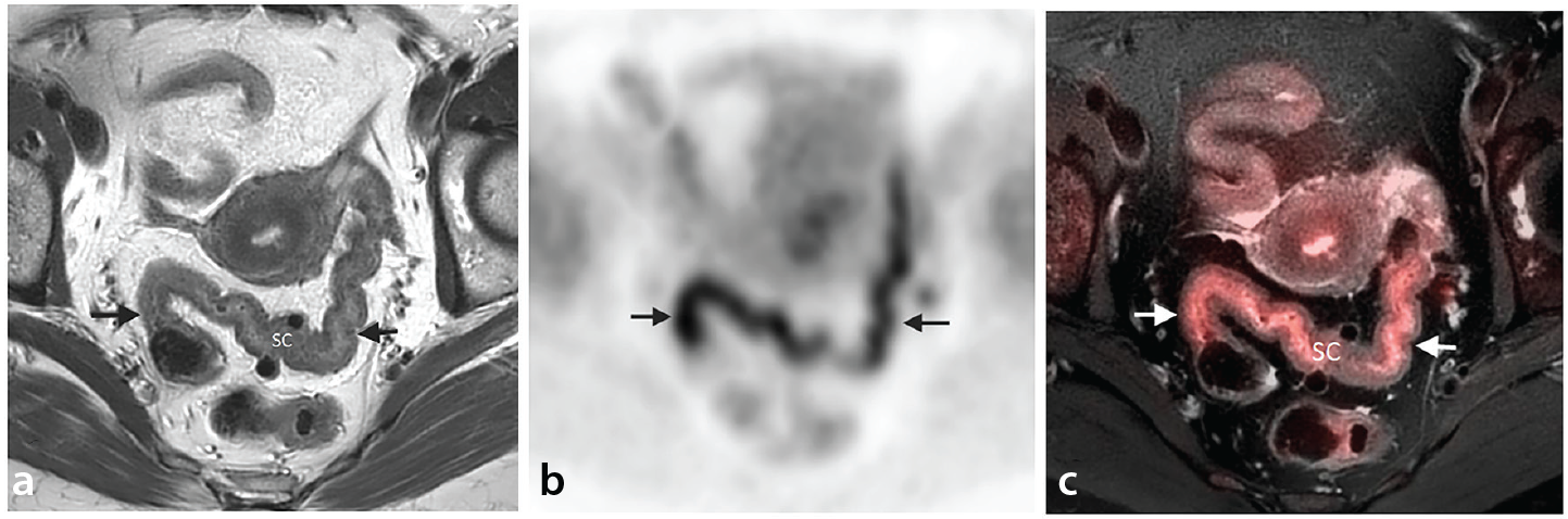
Effect of metformin on intestinal fluorodeoxyglucose uptake
Metformin, a widely used oral antidiabetic, is known to increase FDG uptake in the intestines, especially in the sigmoid colon and rectum (). Although the mechanism is not fully understood, this effect may mimic pathological lesions and result in false-positive findings during oncologic evaluation.
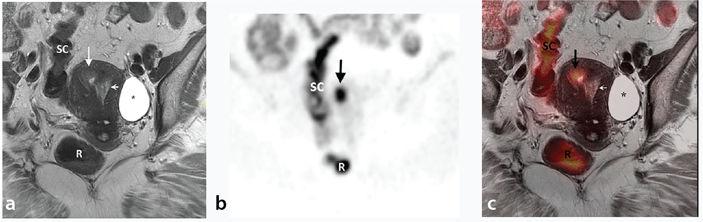
If focal colonic uptake is identified, colonoscopy may be required for further evaluation to rule out neoplasia. Despite this effect of metformin on intestinal FDG uptake, the European Association of Nuclear Medicine guideline (v2.0) recommends continuing this drug unless there are other clinical concerns.
Anal uptake from internal anal sphincter activity
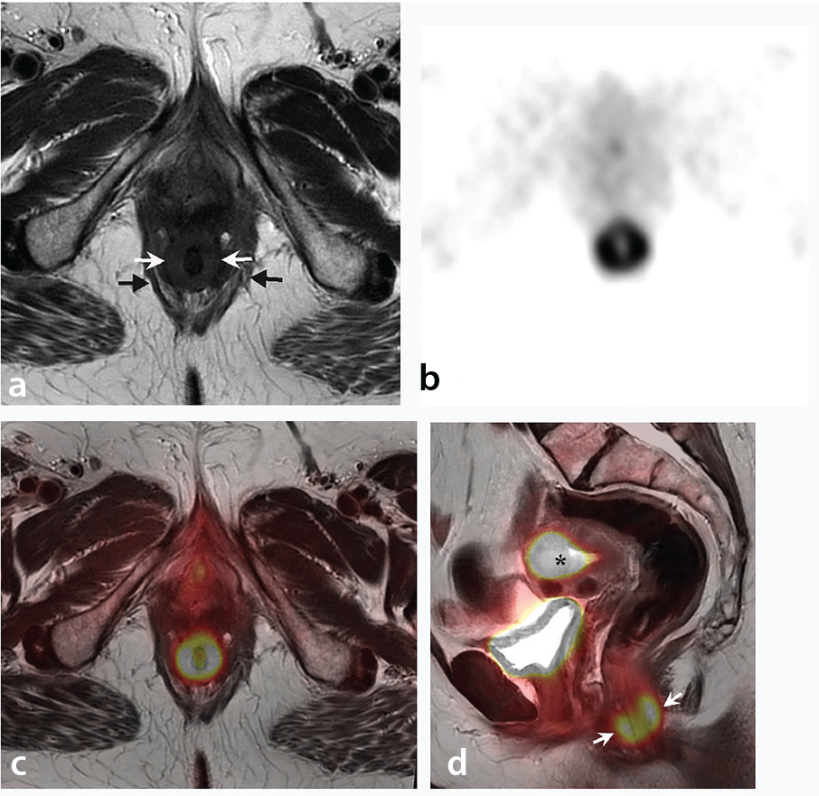
The internal anal sphincter (IAS) is a smooth muscle structure that maintains resting anal tone and is responsible for >70% of baseline anal pressure., Physiological FDG uptake in the IAS is a well-recognized finding on pelvic PET/MRI (), typically seen at its distal end. This uptake is attributed to the basal tone and metabolic activity of the sphincter muscle, even in the absence of anal disease, such as hemorrhoids or fissures.
Muscular fluorodeoxyglucose uptake
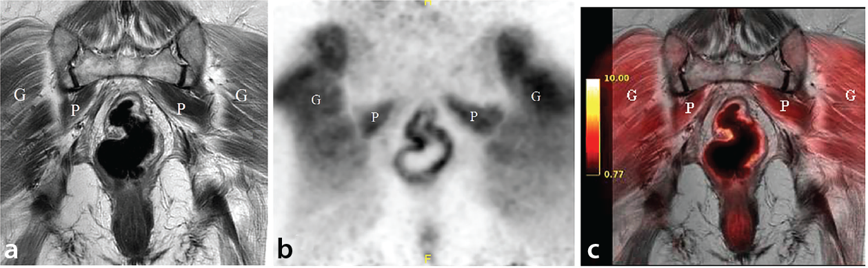
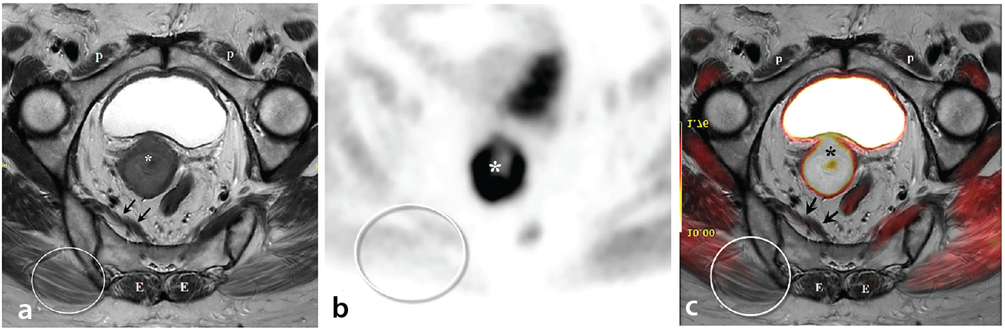
Increased FDG uptake in skeletal muscles can result from voluntary or involuntary contractions. Normally, pelvic muscle uptake is either absent or very mild and homogeneous. Intense uptake may occur due to the following:
• Recent insulin administration,
• Strenuous physical activity within 6 hours of imaging,
• Inadequate fasting (minimum 4 hours required before FDG injection).
Uptake typically appears linear and symmetric (), but may sometimes be focal or asymmetric (), potentially complicating interpretation.
Bone marrow fluorodeoxyglucose uptake
Although there is no physiological FDG uptake in the cortical bone, uptake may be observed in the bone marrow, which may vary depending on patient factors and hematopoietic activity (). Conditions such as anemia, infection, inflammation, or use of hematopoietic stimulants [e.g., granulocyte colony-stimulating factor (G-CSF), erythropoietin] may lead to diffuse bone marrow hypermetabolism., ,
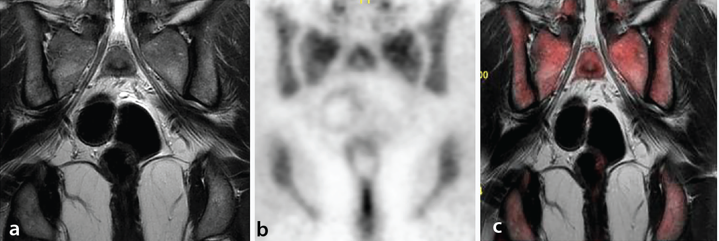
Following administration of pegylated G-CSF, a minimum interval of 3 weeks is recommended before performing PET imaging to avoid false-positive marrow uptake.
Benign Gynecologic Lesions
Endometritis refers to inflammation or infection of the endometrial lining. On MRI, it may present as endometrial thickening with heterogeneous signal intensity, which is non-specific. The differential diagnosis includes endometrial carcinoma, intrauterine hematoma, and endometrial polyps.
FDG PET/MRI typically shows faint FDG uptake in endometritis (), helping to distinguish it from endometrial cancer. Minimal metabolic activity supports a benign etiology.
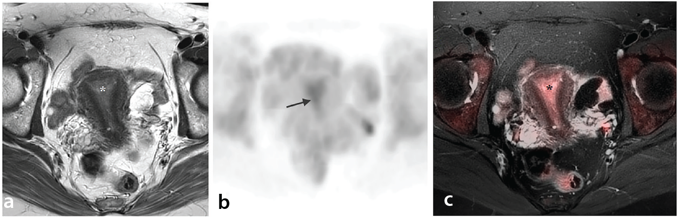
Adenomyosis is a benign condition characterized by ectopic endometrial glands within the myometrium. In premenopausal women, adenomyosis often demonstrates mild FDG uptake (), which may increase during menstruation or ovulation.,
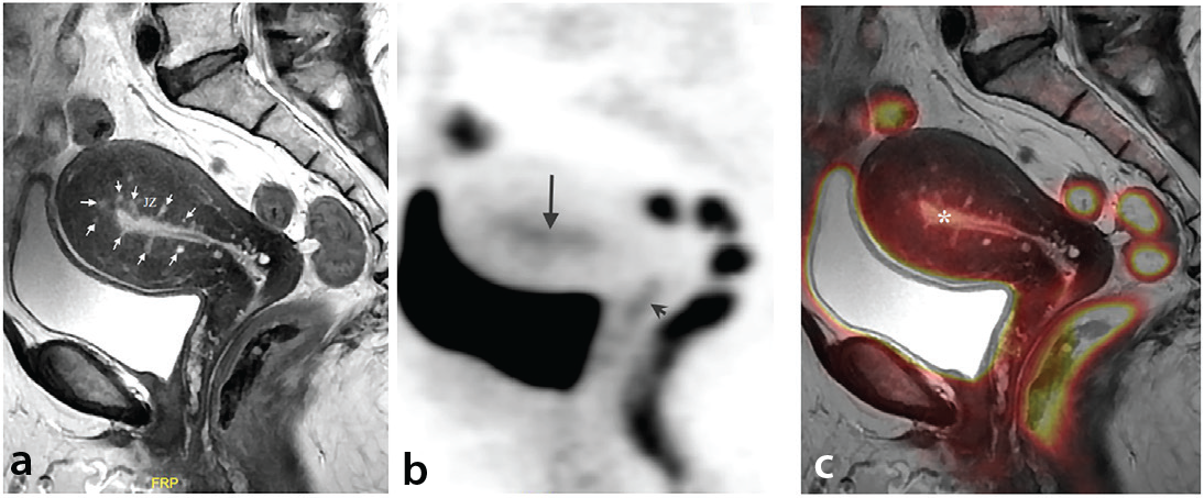
In patients with gynecologic malignancies, FDG-avid adenomyosis may mimic or obscure intrauterine metastases. The MRI component of PET/MR is highly valuable, typically revealing a globally enlarged uterus with a thickened junctional zone containing tiny hyperintense foci on T2-weighted imaging ().
Endometrial hyperplasia is defined by an increased gland-to-stroma ratio due to abnormal glandular proliferation. On T2-weighted MRI, it appears as a diffusely thickened endometrium, with low or intermediate signal intensity, though imaging features are non-specific.
FDG uptake in endometrial hyperplasia is typically mild [mean standardized uptake value (SUV) ~2], significantly lower than in endometrial carcinoma (mean SUV ~9.3). It often occurs in peri- or postmenopausal women ().
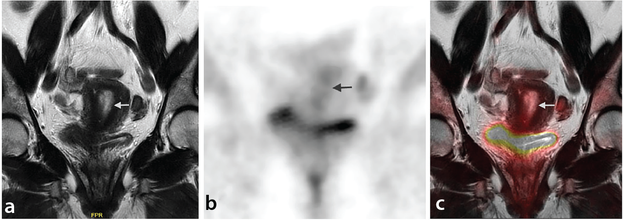
Endometrial polyps are benign overgrowths of endometrial glands and stroma that protrude into the uterine cavity. Although usually indolent, they may carry a risk of malignant transformation.,
On FDG PET, polyps generally exhibit faint or no uptake. However, hypermetabolic activity has been reported, similar to carcinomas. In this study, cases of histopathologically confirmed polyps did not show significant FDG avidity (). In contrast, in a case of serous carcinoma arising within a polyp, the fusion PET/MR image clearly demonstrated marked FDG uptake in the cancerous portion while the polyp itself remained photopenic ().
Leiomyomas (fibroids) are the most common benign gynecologic tumors. FDG uptake is uncommon, reported in approximately 10.4% of premenopausal and 1.2% of postmenopausal women. Uptake, when present, is typically mild (), although rare cases with high FDG avidity exist, particularly during the proliferative phase of the menstrual cycle.,
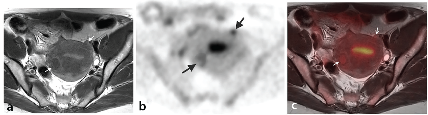
Uptake variability may be influenced by cellularity, vascularity, hormonal status, or degenerative changes. MRI remains essential for characterization and differentiation from malignancy (). Importantly, new FDG avidity in a known leiomyoma does not necessarily indicate malignant transformation.
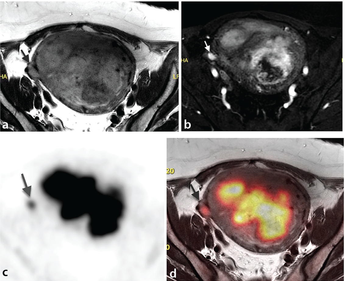
Smooth muscle tumors of uncertain malignant potential lesions exhibit histologic features that overlap with leiomyosarcoma, but with much less aggressive behavior. These rare tumors can demonstrate intense FDG uptake and may coexist with benign leiomyomas ().
Vaginal condyloma acuminata (genital warts) is a common sexually transmitted infection caused by low-risk types of human papillomavirus. In patients undergoing PET/MRI for gynecologic malignancies, vaginal FDG uptake due to condylomas may raise suspicion for metastasis (). Awareness of this entity is crucial to avoid false-positive interpretations.
Bartholin gland cysts result from ductal obstruction and are typically seen in women of reproductive age. Uninfected cysts do not show FDG uptake. However, when infected or forming an abscess, they may become FDG-avid (), often appearing as a small focus in the distal vagina.
Posttreatment Changes in Pelvic Tissues
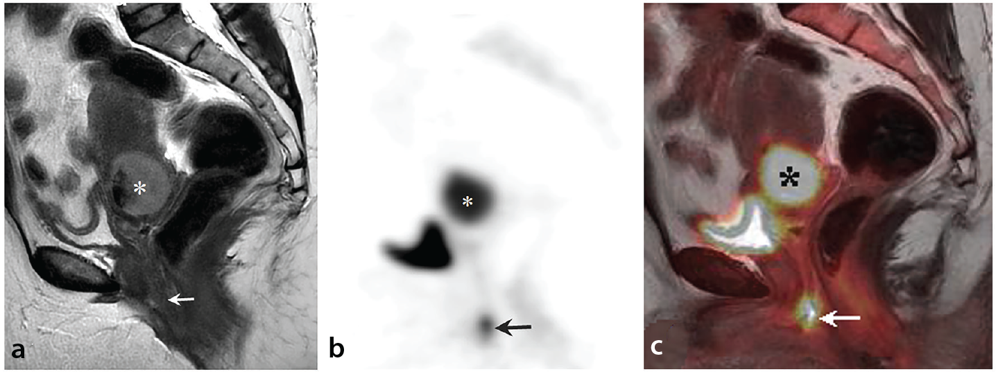
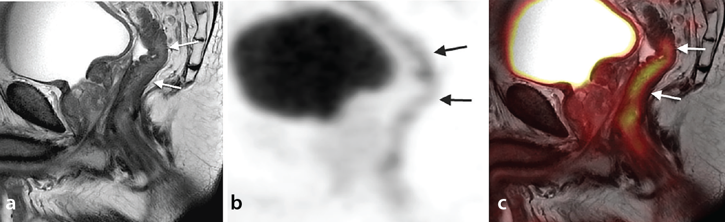
FDG uptake in the presacral region following rectal cancer surgery may raise concern for local recurrence. However, benign postoperative complications, such as anastomotic leakage, can result in inflammatory changes, abscesses, or sinuses, which may also exhibit FDG avidity.
Studies have reported a positive predictive value of only 58% for FDG-avid presacral lesions in identifying true local recurrence. The high soft-tissue contrast of the MRI component of PET/MRI helps differentiate between recurrent tumor and postoperative fibrosis. In such cases, diffusion-weighted imaging (DWI) is reported to be helpful for further characterization. However, it can sometimes be difficult to comment on the nature of lesions that are of intermediate intensity on T2-weighted images and FDG-avid on PET ().
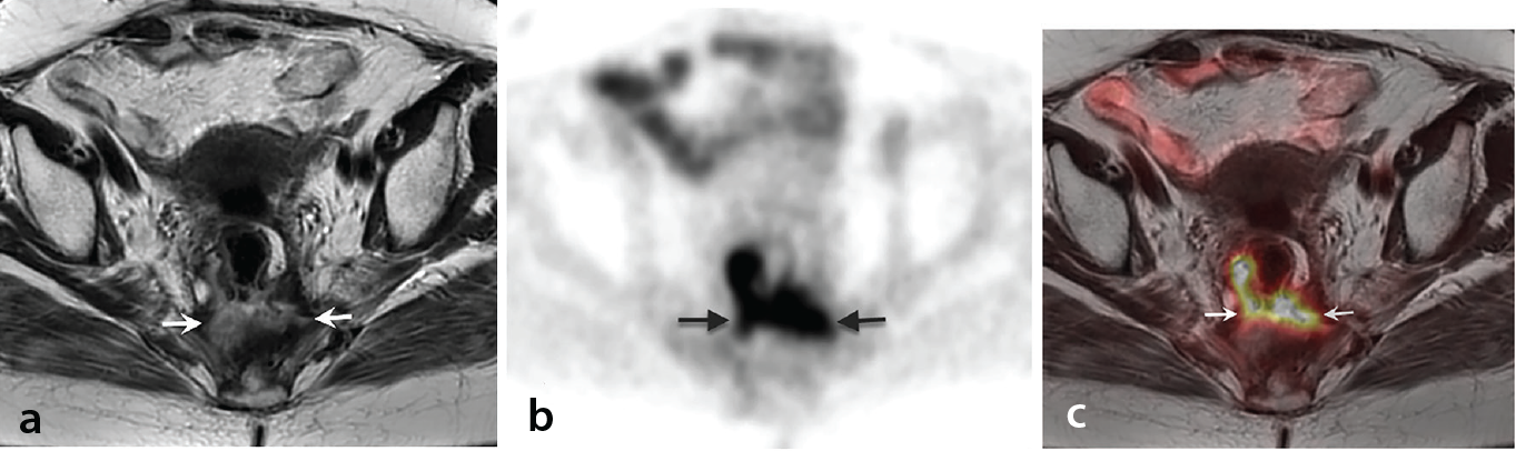
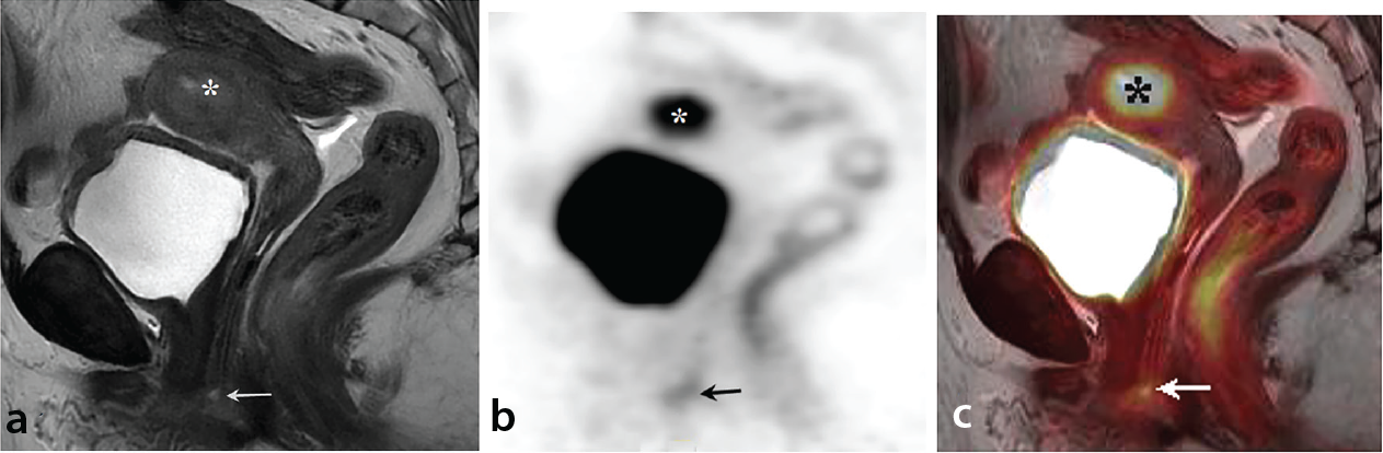
Radiation-induced proctitis, inflammation of the rectal mucosa, is a complication of pelvic radiotherapy, particularly in patients with cervical, rectal, and prostate cancer. Early imaging findings on MRI include submucosal edema and high signal intensity of the rectal wall on T2-weighted images, along with prominent mucosal enhancement. Progressive damage leads to wall thickening and elevated signal in the muscularis layer.,
Mild FDG uptake can be observed in the irradiated rectum on PET/MRI (). Although this generally does not interfere with post-treatment response assessment in patients with rectal cancer, it may complicate interpretation in cases with only partial metabolic response, as simultaneous inflammatory activity cannot be fully excluded.
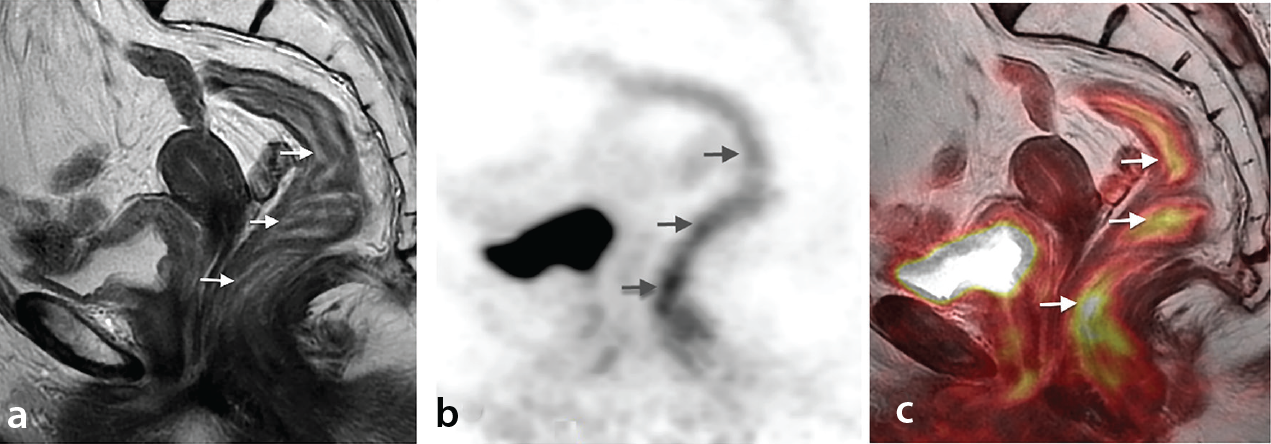
Fat necrosis is a benign process that typically occurs after surgery, trauma, or infection. In this entity, a fibrous connective tissue capsule covers necrotic and degenerated fatty tissue. On imaging, it can mimic tumor recurrence by appearing as a mass-like lesion. Features in MRI often include a well-circumscribed lesion with a hyperintense fatty center and a hypointense fibrous capsule–the so-called “doughnut sign.”
Increased FDG uptake may be observed along the periphery of fat necrosis lesions (), and this uptake can persist for up to 12 months postoperatively. Importantly, serial imaging typically shows no change in lesion size or metabolic activity, supporting a benign etiology.
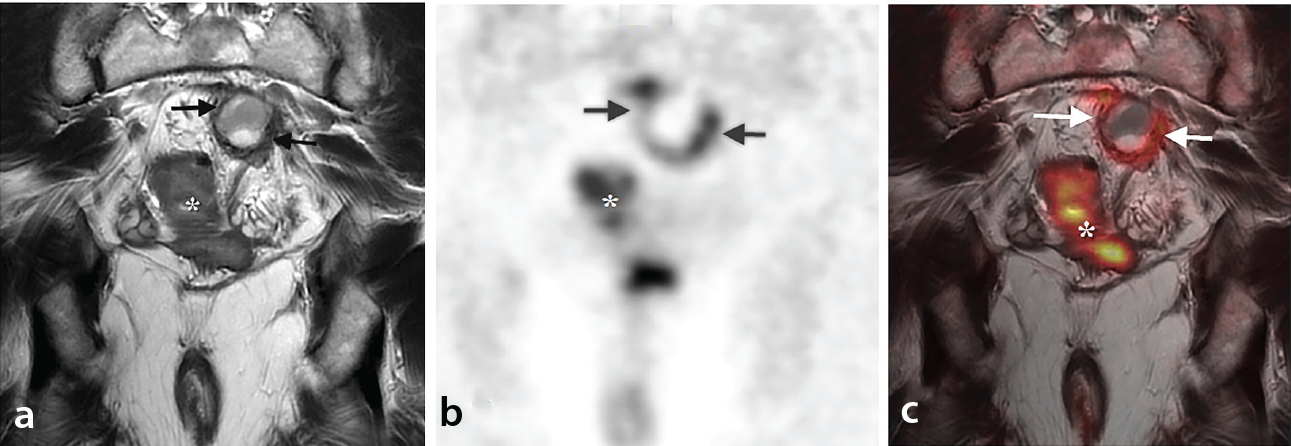
Radiation-induced changes in the sacral bone marrow are a common consequence of pelvic radiotherapy. These include radiation osteitis, insufficiency fractures, and, less commonly, osteoradionecrosis. These changes may present with sacral FDG uptake on PET imaging ().
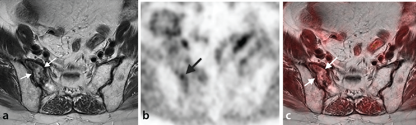
Differentiating between benign post-radiation effects and metastatic bone lesions is essential for accurate interpretation. The MRI component of the PET/MRI study can be useful in localizing and characterizing foci of increased FDG accumulation within the bone. MRI can reveal these changes bilaterally or unilaterally, with findings more prominent adjacent to the sacroiliac joints.
Various Infectious and Inflammatory Lesions
General mechanism of fluorodeoxyglucose uptake in inflammation and infection
FDG uptake in infection and inflammation is mediated by activated inflammatory cells, particularly neutrophils and cells of the monocyte/macrophage lineage. These cells demonstrate upregulated glucose transporter (GLUT) expression (mainly GLUT 1 and GLUT3) and enhanced hexokinase activity, leading to increased glucose metabolism. Both acute and chronic inflammation can produce intense FDG uptake.
Inflammatory bowel disease includes Crohn’s disease and ulcerative colitis, each with distinct imaging patterns. PET/MRI offers a non-invasive method for assessing disease extent and activity. Increased FDG uptake may appear as focal or linear enhancement along affected bowel segments (). Additionally, PET imaging may be helpful in therapy monitoring and in distinguishing between fibrotic and active inflammatory strictures.,
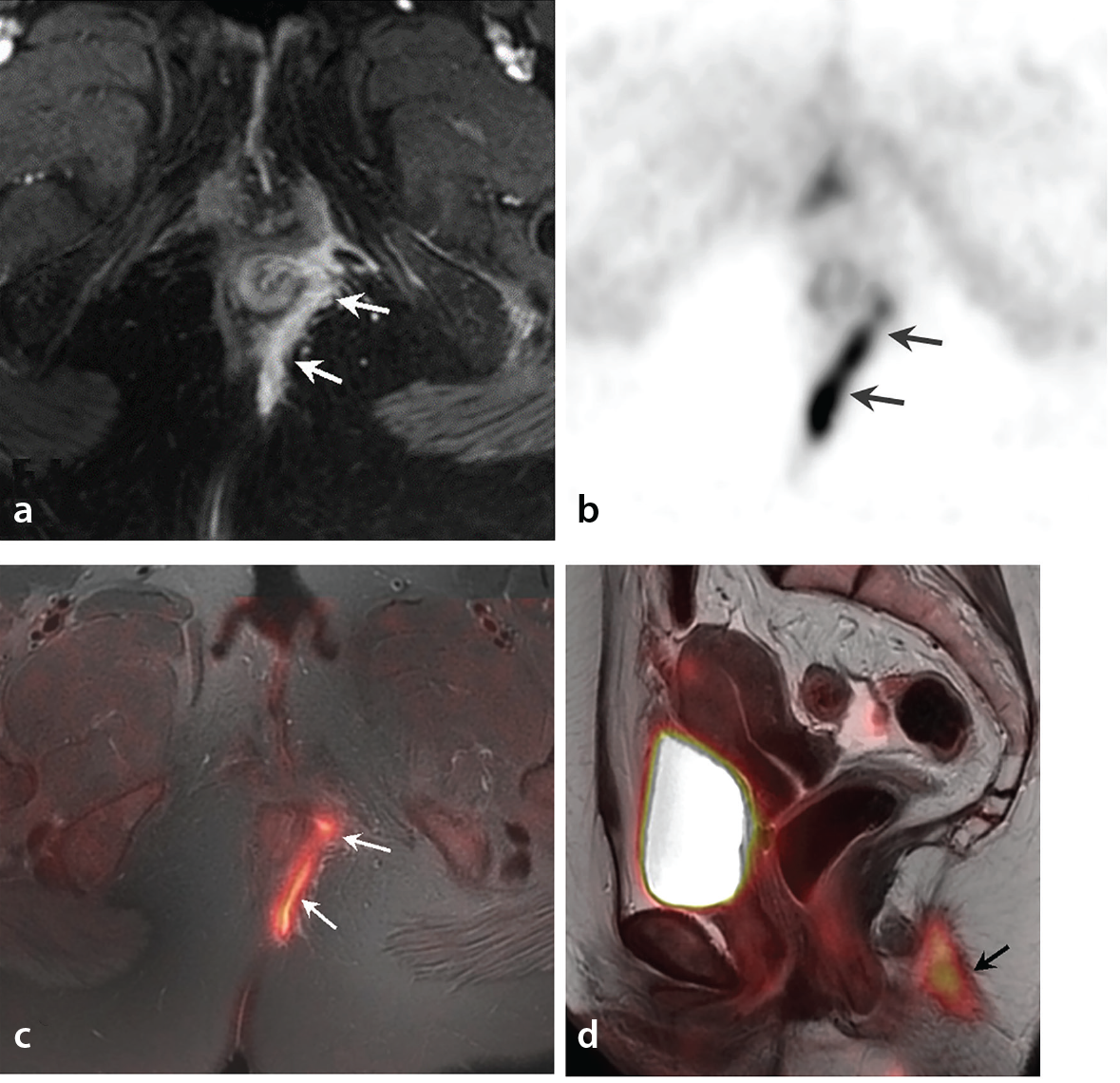
Perianal fistula is an infected tract that develops between the anal canal and the perianal skin, often resulting from prior or ongoing abscess formation. FDG uptake in perianal fistulas may be encountered incidentally in PET/MRIs obtained for oncological purposes ().
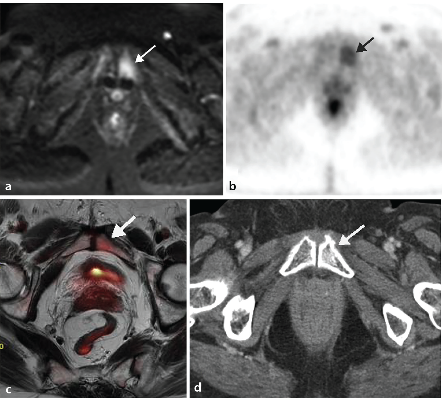
Osteitis pubis is a non-infectious inflammatory condition affecting the pubic symphysis and surrounding structures. It has been associated with pelvic surgery, childbirth, trauma, urological interventions, and repetitive mechanical stress.
MRI may demonstrate parasymphyseal bone marrow edema, diffusion restriction (), and surrounding soft-tissue inflammation. On PET/MRI, moderate to intense unilateral or bilateral FDG uptake near the symphysis pubis is observed ().
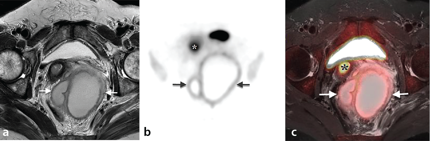
Pelvic abscess formation may result from surgical complications or underlying inflammatory conditions, such as pelvic inflammatory disease, diverticulitis, or inflammatory bowel disease. Imaging reveals complex cystic masses with heterogeneous contents, thick enhancing walls, and perilesional fat stranding. Typically, FDG PET shows intense peripheral uptake with a centrally photopenic (non-avid) core, which is characteristic of abscesses ().
In conclusion, PET/MRI is a powerful imaging modality that integrates the functional capabilities of FDG PET with the superior anatomical detail and tissue characterization provided by MRI. A comprehensive understanding of physiological FDG uptake patterns–in the uterus, fallopian tubes, ovaries, bowel, muscles, and bone marrow–is essential to avoid misinterpretation. Moreover, recognizing benign conditions and posttreatment changes that may mimic malignancy helps reduce false-positive findings and unnecessary interventions.
The MRI component of PET/MRI adds significant value by offering high-resolution anatomical information, functional insights (by DWI), lesion characterization, and reduced radiation exposure compared with PET/CT. These advantages make PET/MRI a highly valuable tool in oncologic imaging, particularly in complex cases involving the pelvis.
Footnotes
Conflict of interest disclosure
The authors declared no conflicts of interest.
References
Lerman H, Metser U, Grisaru D, Fishman A, Lievshitz G, Even-Sapir E. Normal and abnormal 18F-FDG endometrial and ovarian uptake in pre- and postmenopausal patients: assessment by PET/CT. J Nucl Med. 2004;45(2):266-271.
Yu JI, Huh SJ, Kim YI, Kim TJ, Park BK. Variable uterine uptake of FDG in adenomyosis during concurrent chemoradiation therapy for cervical cancer. Radiat Oncol J. 2011;29(3):214-217.
Yun M, Cho A, Lee JH, Choi YJ, Lee JD, Kim CK. Physiologic 18F-FDG uptake in the fallopian tubes at mid cycle on PET/CT. J Nucl Med. 2010;51(5):682-685.
Bonde AA, Korngold EK, Foster BR, et al. Radiological appearances of corpus luteum cysts and their imaging mimics. Abdom Radiol (NY). 2016;41(11):2270-2282.
Ames J, Blodgett T, Meltzer C. 18F-FDG uptake in an ovary containing a hemorrhagic corpus luteal cyst: false-positive PET/CT in a patient with cervical carcinoma. AJR Am J Roentgenol. 2005;185(4):1057-1059.
Lakhani A, Khan SR, Bharwani N, et al. FDG PET/CT Pitfalls in gynecologic and genitourinary oncologic ımaging. Radiographics. 2017;37(2):577-594.
Gontier E, Fourme E, Wartski M, et al. High and typical 18F-FDG bowel uptake in patients treated with metformin. Eur J Nucl Med Mol Imaging. 2008;35(1):95-99.
Boellaard R, Delgado-Bolton R, Oyen WJ, et al. FDG PET/CT: EANM procedure guidelines for tumour imaging: version 2.0. Eur J Nucl Med Mol Imaging. 2015;42(2):328-354.
Keef KD, Cobine CA. Control of motility in the internal anal sphincter. J Neurogastroenterol Motil. 2019;25(2):189-204.
Kumar L, Emmanuel A. Internal anal sphincter: clinical perspective. Surgeon. 2017;15(4):211-226.
Parida GK, Roy SG, Kumar R. FDG-PET/CT in skeletal muscle: pitfalls and pathologies. Semin Nucl Med. 2017;47(4):362-372.
Minamimoto R, Baratto L, Iagaru A. Association between time since administration of pegylated G-CSF (Pegfilgrastim) and bone marrow uptake on FDG PET/CT: determination of a minimum interval. AJR Am J Roentgenol. 2022;218(2):351-358.
Blodgett TM, Ames JT, Torok FS, McCook BM, Meltzer CC. Diffuse bone marrow uptake on whole-body F-18 fluorodeoxyglucose positron emission tomography in a patient taking recombinant erythropoietin. Clin Nucl Med. 2004;29(3):161-163.
Pintican R, Bura V, Zerunian M, et al. MRI of the endometrium - from normal appearances to rare pathology. Br J Radiol. 2021;94(1125):20201347.
Kitajima K, Murakami K, Kaji Y, Sugimura K. Spectrum of FDG PET/CT findings of uterine tumors. AJR Am J Roentgenol. 2010;195(3):737-743.
Sebastiano J, Rodriguez C, Samuels ZV, Pepin K, Zeglis BM. Molecular Imaging in Gynecology: Beyond Cancer. J Nucl Med. 2024;65(7):998-1003.
Tsujikawa T, Okazawa H, Yoshida Y, et al. Distinctive FDG and FES accumulation pattern of two tamoxifen-treated patients with endometrial hyperplasia. Ann Nucl Med. 2008;22(1):73-77.
Kawaguchi M, Kato H, Suzui N, et al. MR imaging findings differentiating uterine submucosal polypoid adenomyomas from endometrial polyps. Br J Radiol. 2019;92(1095):20180430.
Salem AE, Fine GC, Covington MF, et al. PET-CT in Clinical Adult Oncology-IV. Gynecologic and Genitourinary Malignancies. Cancers (Basel). 2022;14(12):3000.
Nishizawa S, Inubushi M, Kido A, et al. Incidence and characteristics of uterine leiomyomas with FDG uptake. Ann Nucl Med. 2008;22(9):803-810.
Kitajima K, Murakami K, Yamasaki E, Kaji Y, Sugimura K. Standardized uptake values of uterine leiomyoma with 18F-FDG PET/CT: variation with age, size, degeneration, and contrast enhancement on MRI. Ann Nucl Med. 2008;22(6):505-512.
Zhang R, Tian X, Qin L, Lu D, Shen J. High 18F-FDG uptake for uterine smooth muscle tumor of uncertain malignant potential. Clin Nucl Med. 2015;40(4):349-351.
Kishimoto T, Mabuchi S, Kato H, Kimura T. Condyloma acuminata induces focal intense FDG uptake mimicking vaginal stump recurrence from uterine cervical cancer: a case report. Eur J Gynaecol Oncol. 2013;34(1):99-100.
Dejanovic D, Hansen NL, Loft A. PET/CT Variants and pitfalls in gynecological cancers. Semin Nucl Med. 2021;51(6):593-610.
Pennings JP, de Haas RJ, de Jong KP, Dierckx RAJO, Kwee TC. Beware of false-positive FDG PET/CT interpretations for presacral recurrent rectal cancer. Clin Nucl Med. 2019;44(5):e342-e344.
Pennings JP, de Haas RJ, Murshid KJA, de Jong KP, Dierckx RAJO, Kwee TC. FDG-avid presacral soft tissue mass in previously treated rectal cancer: diagnostic outcome and additional value of MRI, including diffusion-weighted imaging. Eur J Surg Oncol. 2019;45(4):606-612.
Kalff V, Ware R, Heriot A, Chao M, Drummond E, Hicks RJ. Radiation changes do not interfere with postchemoradiation restaging of patients with rectal cancer by FDG PET/CT before curative surgical therapy. Int J Radiat Oncol Biol Phys. 2009;74(1):60-66.
Kalff V, Duong C, Drummond EG, Matthews JP, Hicks RJ. Findings on 18F-FDG PET scans after neoadjuvant chemoradiation provides prognostic stratification in patients with locally advanced rectal carcinoma subsequently treated by radical surgery. J Nucl Med. 2006;47(1):14-22.
Davidson T, Lotan E, Klang E, et al. Fat necrosis after abdominal surgery: a pitfall in interpretation of FDG-PET/CT. Eur Radiol. 2018;28(6):2264-2272.
Meixel AJ, Hauswald H, Delorme S, Jobke B. From radiation osteitis to osteoradionecrosis: incidence and MR morphology of radiation-induced sacral pathologies following pelvic radiotherapy. Eur Radiol. 2018;28(8):3550-3559.
Abikhzer G, Treglia G, Pelletier-Galarneau M, et al. EANM/SNMMI guideline/procedure standard for [ 18 F]FDG hybrid PET use in infection and inflammation in adults v2.0. Eur J Nucl Med Mol Imaging. 2025;52(2):510-538.
Lovinfosse P, Hustinx R. The role of PET imaging in inflammatory bowel diseases: state-of-the-art review. Q J Nucl Med Mol Imaging. 2022;66(3):206-217.
Broos WAM, van der Zant FM, Knol RJJ. Athletic osteitis pubis detected on 18 F-FDG PET/CT. Clin Nucl Med. 2022;47(9):822-823.