
Dear Editor,
We read with interest the review article by Stanietzky et al. entitled “Unusual liver tumors: spectrum of imaging findings with pathologic correlation” in the recent issue of Diagnostic and Interventional Radiology. In this article, the authors provide both textual and visual portrayals of hepatic neoplasms that are both unusual and rare. The authors also discussed the subject of “solitary plasmacytoma” under the subtitle of “Multiple myeloma and solitary plasmacytoma” and stated that this rare lesion has variable imaging findings. In this context, we would like to share the liver imaging findings of our case that was histopathologically diagnosed as “hepatic solitary plasmacytoma.”
A 50-year-old woman presented with diffuse abdominal pain that had intensified at the right upper quadrant for around 3 months. Aside from laparoscopic cholecystectomy, her medical history was unremarkable, and her physical examination was normal. Hemogram, liver function, and renal function test results were in normal range. Erythrocyte sedimentation rate was 33 mm/h (normal: 0–25 mm/h). Viral hepatitis markers and tumor markers (alpha fetoprotein, CEA, CA 15-3, CA19-9, CA125) were negative.
The patient subsequently underwent contrast-enhanced dynamic computed tomography (CT) of the liver. A hypervascular hepatic mass at segment 2 that showed progressive washout was detected on CT (). For further characterization of the hepatic lesion, magnetic resonance imaging of the upper abdomen was performed using a 3.0-T system, which showed a solid hepatic mass with thin septations and irregular lobulated borders, measuring 4.5 × 3.5 cm at segment 2. The lesion was hypointense on T1-weighted images and hyperintense relative to the liver on T2-weighted images (). The lesion remained hyperintense on the diffusion weighted images obtained, with a b-value of 1,000 s/mm2. It was slightly hyperintense on the apparent diffusion coefficient map, except for a small hypointense component located at its lower part (, , ). On dynamic imaging performed following intravenous injection of a hepatospecific contrast agent, the mass showed early enhancement in the arterial phase and progressive wash-out in the venous phases (, ). The lesion was hypointense relative to the liver on hepatobiliary phase ().
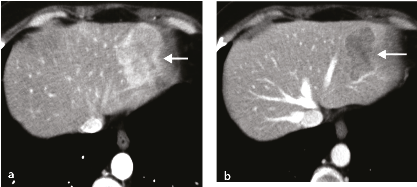
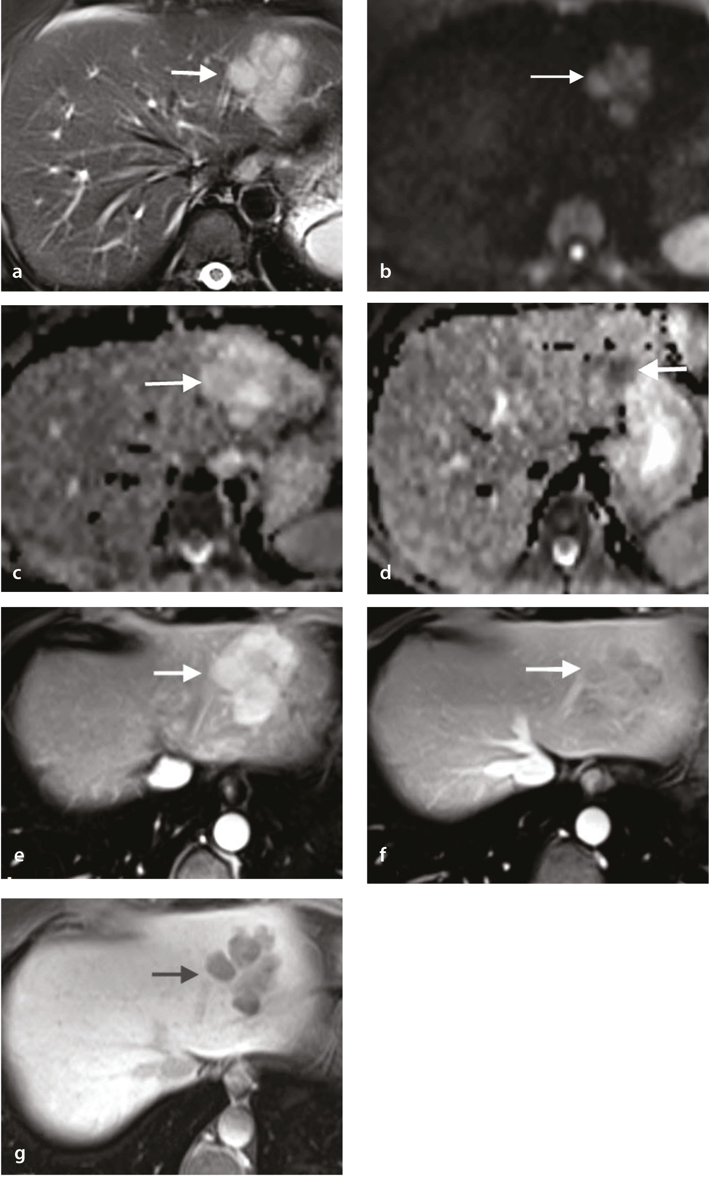
Based on the imaging findings, the differential diagnosis included hypervascular liver lesions. Hepatocellular carcinoma was not considered the primary diagnosis due to the absence of a history of chronic liver disease. The lesion’s morphology and hepatobiliary phase images were not typical for focal nodular hyperplasia. The lack of intralesional fat made the diagnosis of hepatic adenoma less likely. As the imaging findings were non-specific and inconclusive, and a possible hypervascular metastasis could not be ruled out, a liver biopsy was deemed appropriate.
Ultrasound-guided fine needle aspiration biopsy result was compatible with plasmacytoma (). The definitive diagnosis was made via immunocytochemical analysis of cell block material. Immunocytochemical study showed that tumor cells were strongly positive for CD38 and CD138 antibodies and displayed lambda monoclonality.
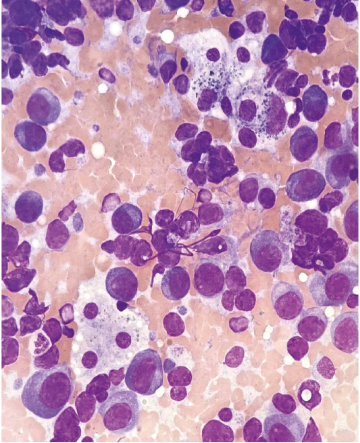
Serum electrophoresis showed immunoglobulin G (IgG) monoclonal gammopathy. The free light chain assay ratio was abnormal (free kappa/free lambda ratio: 0.25; normal, 0.26–1.65). The immunofixation of the serum showed IgG lambda monoclonal gammopathy. Concentrated urine immunofixation (×50) revealed the presence of lambda light chain.
The imaging findings of the entity known as “solitary plasmacytoma” are nonspecific.,, Based on our observations, it can be stated that, in the relevant clinical setting, it would be appropriate to keep the possibility of plasmacytoma in mind in the differential diagnosis of an arterially enhancing liver mass.
Footnotes
Conflict of interest disclosure
The authors declared no conflicts of interest.
References
Stanietzky N, Salem AE, Elsayes KM, et al. Unusual liver tumors: spectrum of imaging findings with pathologic correlation. Diagn Interv Radiol. 2025;31(2):58-67.
Ooi GC, Chim JC, Au WY, Khong PL. Radiologic manifestations of primary solitary extramedullary and multiple solitary plasmacytomas. AJR Am J Roentgenol. 2006;186:821-827.
Ng P, Slater S, Radvan G, Price A. Hepatic plasmacytomas: case report and review of imaging features. Australas Radiol. 1999;43(1):98-101.
Ueda K, Matsui H, Watanabe T, et al. Spontaneous rupture of liver plasmacytoma mimicking hepatocellular carcinoma. Intern Med. 2010;49(7):653-657.