
Abstract
Desmoid tumors (DTs) are rare, locally aggressive soft tissue neoplasms with highly variable clinical behavior. Although benign, their infiltrative nature can lead to considerable morbidity. DTs present a major challenge due to their unpredictable behavior and potential for misdiagnosis. Recently, there has been a large shift in the treatment strategy for DTs, and the number of cases being followed up with imaging has increased. Cryoablation has emerged as a minimally invasive treatment option, yet post-procedural imaging features remain poorly characterized. This study illustrates the magnetic resonance imaging (MRI) evolution of DTs following percutaneous cryoablation, emphasizing key patterns across pre-treatment, intra-procedural, and follow-up studies. The available MRI follow-up ranged from 6 to 30 months. Through a pictorial approach, we aimed to provide radiologists with practical insights to avoid misinterpretation of expected post-treatment changes as recurrence and to improve clinical management.
Keywords: Desmoid tumor, magnetic resonance imaging, follow-up studies, soft tissue neoplasm, ablation technique, cryoablation, radiology, interventional
Main points
• Magnetic resonance imaging (MRI) is fundamental in the post-cryoablation follow-up of desmoid tumors, enabling precise differentiation between expected treatment-related changes and tumor recurrence.
• Key imaging findings after cryoablation include a central non-enhancing cavity, peripheral granulation tissue with contrast enhancement, and progressive fibrosis reflected by decreasing T2 signal intensity.
• Recognizing the temporal evolution of these MRI features is essential to avoid misinterpretation and to guide appropriate clinical decision-making.
Desmoid-type fibromatosis, also known as desmoid tumor (DT) or aggressive fibromatosis, is a rare, locally invasive fibroblastic proliferation characterized by an unpredictable clinical course., These tumors account for < 3% of soft tissue neoplasms, with an incidence of approximately 3–5 cases per million annually. They typically affect individuals in their third to fourth decades of life and show a higher prevalence in women. Most cases are sporadic and arise in extra-abdominal locations, particularly the extremities and trunk.
DTs pose a major diagnostic and management challenge due to their variable behavior, which can range from rapid growth to spontaneous regression. Recent guidelines emphasize an initial surveillance strategy to prevent overtreatment, reserving intervention for symptomatic or progressive cases. However, a decision to initiate therapy earlier may be considered when DTs are located near critical structures, as there may be a higher risk of morbidity before the disease stabilizes. When treatment is warranted, options include systemic therapies (such as chemotherapy or tyrosine kinase inhibitors) and local interventions.,
Cryoablation has emerged as a promising local therapy and is now included in updated management guidelines. This minimally invasive technique employs cryoprobes to induce tumor cell death through extreme cold, achieved via the Joule–Thomson effect. Cellular destruction results from intracellular ice formation and vascular injury, leading to ischemia. Image-guided cryoablation, typically performed under computed tomography guidance, allows precise visualization of a low-attenuation ice ball in soft tissues, providing visual confirmation that the tumor is included in the cryoablated volume. Recent studies have shown that image-guided percutaneous cryoablation appears to be safe and effective for local control in patients with extra-abdominal DTs.,,,, In this pictorial essay, we illustrate six representative cases of extra-abdominal DTs treated with percutaneous cryoablation, including three men and three women (age range 33–54 years). Tumor sites comprise the gluteal region (n = 2), lumbar wall (n = 1), abdominal wall (n = 1), scapular region (n = 1), and calf (n = 1) ().
Table 1. Patients’ demographics and characteristics
Case | Age (years) | Sex | Tumor location | Magnetic resonance imaging follow-up duration (months) |
|---|---|---|---|---|
1 | 35 | Male | Right gluteal region | 18 |
2 | 54 | Female | Right lumbar subcutaneous region | 30 |
3 | 48 | Female | Left abdominal wall | 8 |
4 | 33 | Male | Left gluteal subcutaneous region | 25 |
5 | 50 | Female | Left scapular region | 18 |
6 | 33 | Female | Calf | 6 |
Role of magnetic resonance imaging
Magnetic resonance imaging (MRI) plays a critical role in the diagnosis, treatment planning, and follow-up of DTs. Imaging findings correlate with histopathologic features; hyperintense areas on T2-weighted and enhancing sequences reflect high cellularity, whereas hypointense, non-enhancing regions correspond to fibrous, low-cellularity components., An increase in collagenization within the tumor is associated with a decrease in cellularity, suggesting a reduction in tumor activity. This can be seen on MRI as a decrease in enhancement and T2 hyperintensity, which are imaging features indicating a positive response to therapy.
Post-cryoablation imaging findings
Post-cryoablation MRI findings evolve. In early follow-up, considerable edema may be noted in the cryoablation area and adjacent soft tissues (). Early imaging signs after the procedure include a non-enhancing ablation cavity with a high T2 signal, corresponding to coagulative necrosis. A thin inner rim of low T2 signal, indicative of hemosiderin rim (hemorrhagic congestion), can be better visualized on T2 gradient echo sequences. A thicker outer rim of intermediate/low T2 signal with post-contrast enhancement corresponds to granulation tissue/vascular fibrosis ().
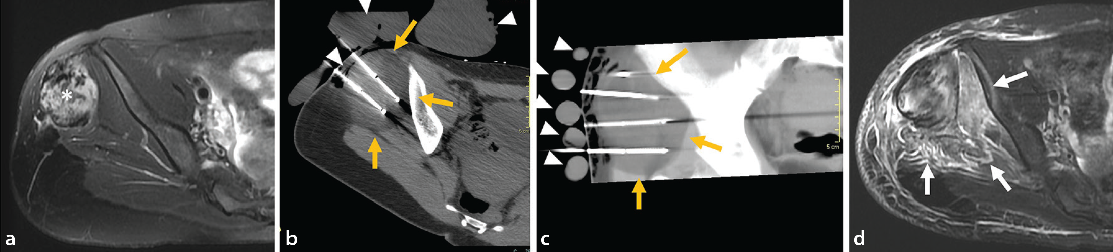
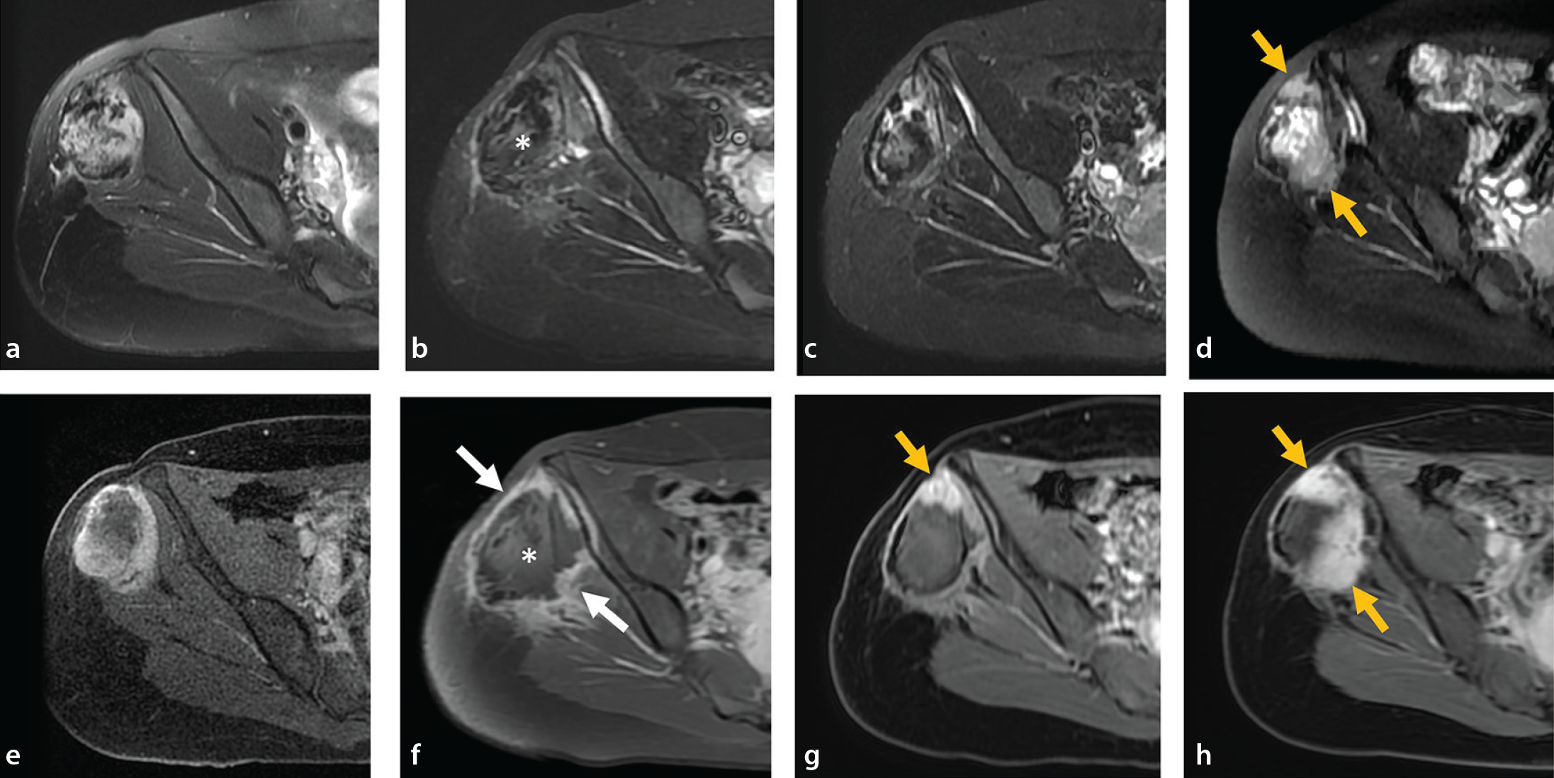
In subsequent follow-ups, the non-enhancing ablation cavity shows a size reduction and may show a marked decrease in T2 signal intensity, indicative of dense fibrosis. Additionally, a progressive reduction in post-contrast enhancement of the outer rim of granulation tissue/vascular fibrosis can be noted (). Finally, a reduction in lesion volume is expected in successful cases (), with a progressive replacement by adipose tissue observed in subcutaneous cases (). If tumor recurrence occurs, it can be observed on post-contrast sequences as a nodular enhancing area (). Imaging findings are summarized in .
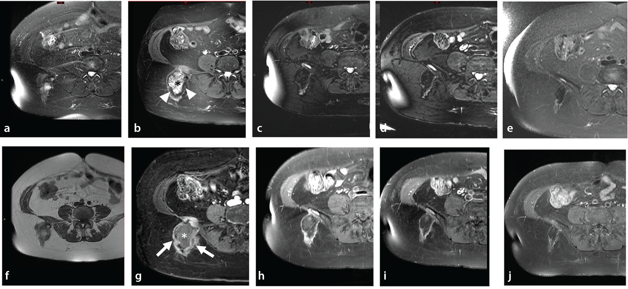
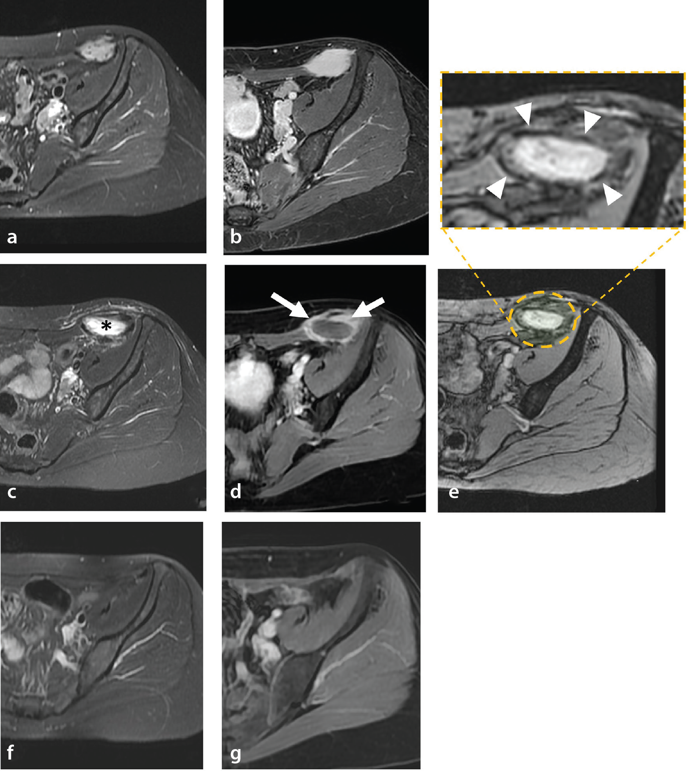
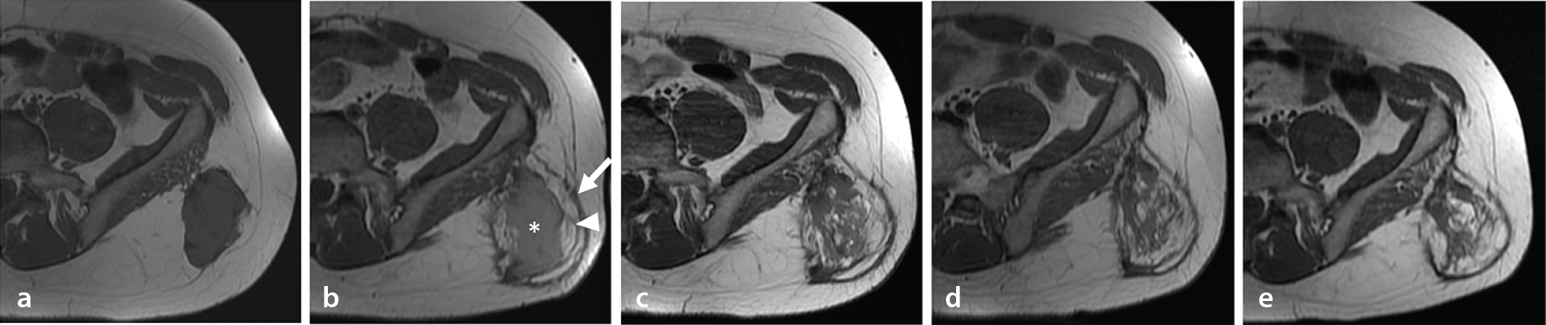
Table 2. Key magnetic resonance imaging findings after cryoablation of desmoid tumors
Imaging feature | Definition/description |
|---|---|
Non-enhancing ablation cavity | Central zone without enhancement, corresponding to coagulative necrosis |
Hemosiderin rim | Thin inner rim of low T2 signal due to hemorrhagic congestion |
Granulation tissue/vascular fibrosis | Outer rim with intermediate/high T2 signal and post-contrast enhancement |
Progressive fibrosis | Reduction in T2 signal and enhancement over time |
Nodular enhancement | New or growing enhancing nodule at cavity margin, suspicious for recurrence |
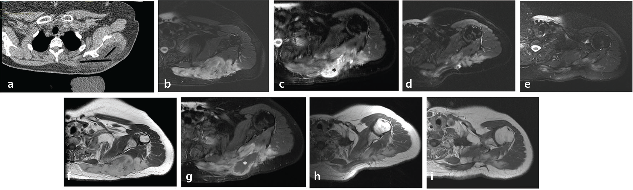
The success rate of cryoablation is directly related to the ice ball encompassing the entire lesion during the procedure. However, cryoablation may induce the “abscopal effect,” leading to the reduction or disappearance of the tumor in non-frozen sites, possibly due to immune modulation (). This phenomenon is of high importance in tumor therapy, as it effectively reduces the likelihood of tumor recurrence.
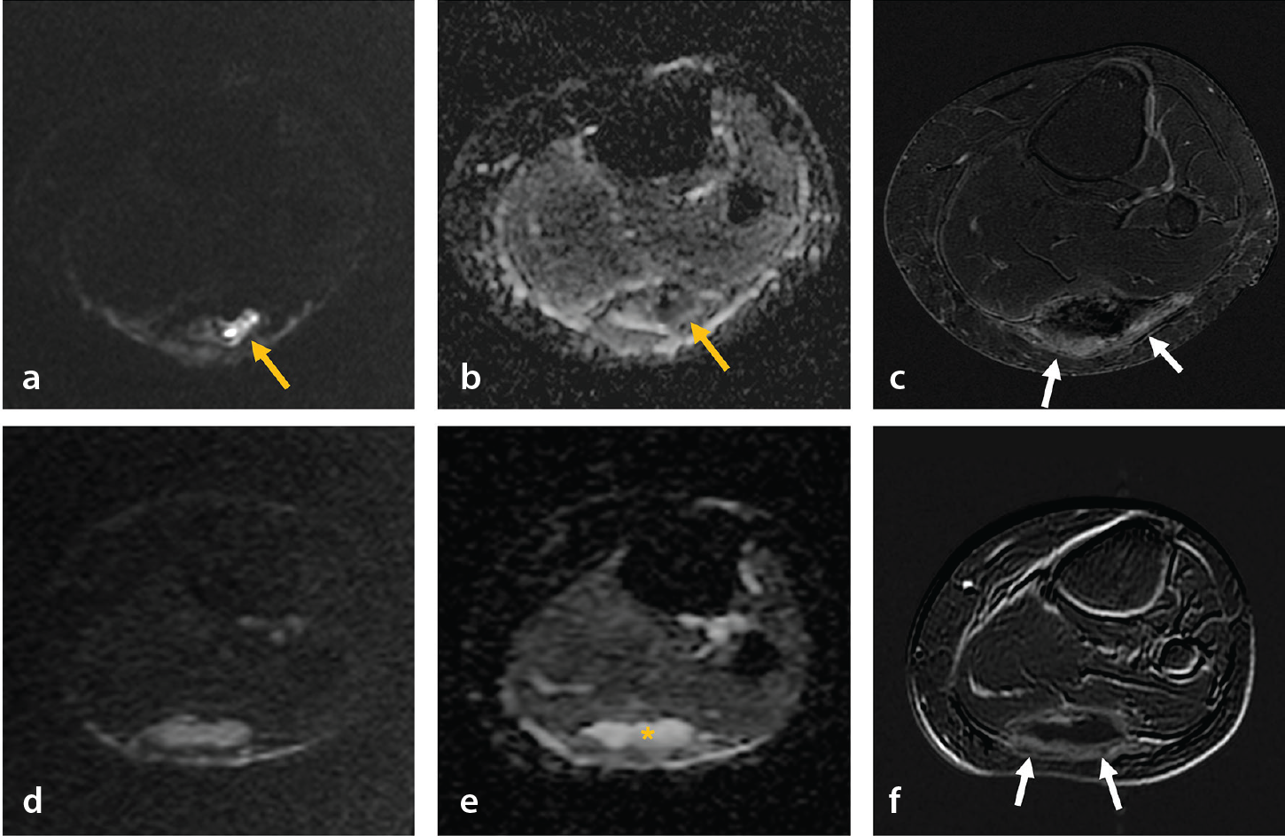
Diffusion-weighted imaging (DWI) correlates with the content of the treated tissue, showing higher apparent diffusion coefficient (ADC) values in cases of low cellularity and fibrous content. It can also be used to detect changes in the treated tissue post-cryotherapy. A recent article reported low ADC values (mean 0.90 × 10³ mm2/s) in early follow-up of treated tissue after cryotherapy, possibly due to tumor necrosis. This should not be confused with recurrence, especially when no enhancement is present. Further studies are needed to confirm DWI patterns in treated tissue post-cryotherapy. However, this pattern was also observed in some cases in this study, as shown in .
Discussion
The management of DTs has become increasingly individualized, tailored around patients’ needs and lesions’ features. Aggressive surgical approaches are generally avoided in favor of more conservative, minimally invasive strategies. The current consensus has shifted towards a “watch-and-wait” first-line approach, and intervention is performed for progressive or symptomatic cases., In this context, image-guided percutaneous cryoablation has emerged as a valuable and minimally invasive alternative to surgery and systemic therapy.
Recent studies have shown encouraging outcomes in terms of local control and symptom relief, especially when complete tumor coverage is achieved during ablation. This, combined with minimal side effects, has led to its inclusion in recent international guidelines., ,, Furthermore, the potential for cryoablation to induce systemic antitumor effects—the so-called abscopal effect—suggests opportunities for synergy with systemic therapies, including immunotherapy.
Despite these advancements, standardized radiologic criteria for treatment response remain lacking. MRI features, such as transient T2 hyperintensity or DWI changes, may mimic recurrence, especially in early follow-up, if not properly contextualized.
A deeper understanding of the temporal evolution of these features is essential to avoid misinterpretation and guide clinical decision-making.
Emerging technologies, including artificial intelligence and radiomics, may offer tools to differentiate residual tumor from post-ablation changes. Recent studies have shown that deep learning applied to baseline MRI can predict clinical progression of DTs with high accuracy (up to 93%), suggesting a potential role for artificial intelligence-based tools in risk stratification and treatment planning. Finally, growing interest in cryoablation’s immunomodulatory potential may support future therapeutic combinations that enhance outcomes in patients with DTs.
In conclusion, our understanding of DTs is evolving rapidly. MRI plays a central role in evaluating post-cryoablation changes and guiding patient management. DTs treated with percutaneous cryoablation exhibit a predictable sequence of imaging changes over time. Key post-treatment MRI findings include a central non-enhancing cavity of coagulative necrosis, peripheral granulation tissue with enhancement, and progressive fibrosis characterized by decreasing T2 signal and enhancement. Accurate recognition of these patterns is critical to distinguish treatment response from recurrence. Familiarity with expected imaging patterns and their timeline can help avoid misdiagnosis of recurrence and optimize treatment strategies. Future research integrating imaging biomarkers, artificial intelligence, and immunologic response will likely further refine our approach to this complex entity.
Footnotes
Conflict of interest disclosure
The authors declared that they have no conflict of interest.
References
Sbaraglia M, Bellan E, Dei Tos AP. The 2020 WHO Classification of Soft Tissue Tumours: news and perspectives. Pathologica. 2021;113(2):70-84.
WHO Classification of Tumours Editorial Board. WHO Classification of Tumours: Soft Tissue and Bone Tumours. 5 th ed. Vol 3 [Internet]. Lyon: International Agency for Research on Cancer; 2020 [cited 2025 Nov 6].
Kasper B, Baumgarten C, Garcia J, et al. An update on the management of sporadic desmoid-type fibromatosis: a European Consensus Initiative between Sarcoma PAtients EuroNet (SPAEN) and European Organization for Research and Treatment of Cancer (EORTC)/Soft Tissue and Bone Sarcoma Group (STBSG). Ann Oncol. 2017;28(10):2399-2408.
Gronchi A, Colombo C, Le Péchoux C, et al. Sporadic desmoid-type fibromatosis: a stepwise approach to a non-metastasising neoplasm–a position paper from the Italian and the French Sarcoma Group. Ann Oncol. 2014;25(3):578-583.
Desmoid Tumor Working Group. The management of desmoid tumours: a joint global consensus-based guideline approach for adult and paediatric patients. Eur J Cancer. 2020;127:96-107.
Papalexis N, Savarese LG, Peta G, et al. The new ice age of musculoskeletal intervention: role of percutaneous cryoablation in bone and soft tissue tumors. Curr Oncol. 2023;30(7):6744-6770.
Auloge P, Garnon J, Robinson JM, et al. Percutaneous cryoablation for advanced and refractory extra-abdominal desmoid tumors. Int J Clin Oncol. 2021;26(6):1147-1158.
Yan YY, Walsh JP, Munk PL, et al. A single-center 10-year retrospective analysis of cryoablation for the management of desmoid tumors. J Vasc Interv Radiol. 2021;32(9):1277-1287.
Efrima B, Ovadia J, Drukman I, et al. Cryo-surgery for symptomatic extra-abdominal desmoids. A proof of concept study. J Surg Oncol. 2021;124(4):627-634.
Saltiel S, Bize PE, Goetti P, et al. Cryoablation of extra-abdominal desmoid tumors: a single-center experience with literature review. Diagnostics (Basel). 2020;10(8):556.
Kurtz JE, Buy X, Deschamps F, et al. CRYODESMO-O1: a prospective, open phase II study of cryoablation in desmoid tumour patients progressing after medical treatment. Eur J Cancer. 2021;143:78-87.
Bouhamama A, Lame F, Mastier C, et al. Local control and analgesic efficacy of percutaneous cryoablation for desmoid tumors. Cardiovasc Intervent Radiol. 2020;43(1):110-119.
Shinagare AB, Ramaiya NH, Jagannathan JP, et al. A to Z of desmoid tumors. AJR Am J Roentgenol. 2011;197(6):W1008-1014.
Ganeshan D, Amini B, Nikolaidis P, Assing M, Vikram R. Current update on desmoid fibromatosis. J Comput Assist Tomogr. 2019;43(1):29-38.
Okuda M, Yoshida K, Kobayashi S, Gabata T. Desmoid-type fibromatosis: imaging features and course. Skeletal Radiol. 2023;52(7):1293-1303.
Chen J, Qian W, Mu F, Niu L, Du D, Xu K. The future of cryoablation: an abscopal effect. Cryobiology. 2020;97:1-4.
Garnon J, Cazzato RL, Autrusseau PA, et al. Desmoid fibromatosis: interventional radiology (sometimes) to the rescue for an atypical disease. Br J Radiol. 2025;98(1170):840-850.
Cazzato RL, Gantzer J, de Marini P, et al. Sporadic desmoid tumours: systematic review with reflection on the role of cryoablation. Cardiovasc Intervent Radiol. 2022;45(5):613-621.
Kasper B, Baldini EH, Bonvalot S, et al. Current management of desmoid tumors: a review. JAMA Oncol. 2024;10(8):1121-1128.
Yang X, Gao X, Xu C, et al. Cryoablation synergizes with anti-PD-1 immunotherapy induces an effective abscopal effect in murine model of cervical cancer. Transl Oncol. 2025;51:102175.
Düx DM, Chodakiewitz Y, Bitton R, et al. MRI features that predict progression of residual disease after ablation of extra-abdominal desmoid fibromatosis. Eur Radiol. 2025;35(7):4161-4170.
Fares R, Atlan LD, Druckmann I, et al. Imaging-based deep learning for predicting desmoid tumor progression. J Imaging. 2024;10(5):122.