
Abstract
Myocardial infarction (MI) is a leading cause of morbidity and mortality worldwide. Although rapid diagnosis and reperfusion in the acute setting rely primarily on clinical assessment, electrocardiography, echocardiography, and invasive coronary angiography, advanced cardiac imaging plays an essential role beyond the hyperacute phase. Cardiac computed tomography (CCT) and cardiac magnetic resonance (CMR) imaging provide complementary information that extends from coronary anatomy to myocardial tissue characterization and functional assessment. CMR imaging enables the comprehensive, multiparametric evaluation of MI, including left ventricular function, myocardial edema and area at risk, infarct size and transmural extent, microvascular obstruction, intramyocardial hemorrhage, myocardial viability, and ischemia using cine imaging, T1/T2 and T2* mapping, perfusion imaging, and late gadolinium enhancement. These features support an accurate differentiation between acute and chronic infarction, an assessment of myocardial salvage, and prognostic stratification. CCT offers a rapid, non-invasive assessment of coronary artery stenosis and plaque characteristics and has expanded to include an evaluation of ventricular function, myocardial perfusion, and delayed-enhancement patterns. When combined with CT-derived fractional flow reserve or myocardial perfusion imaging, CT allows for an integrated anatomic and functional assessment of the myocardium, particularly for non-culprit lesions, following MI. Myocardial delayed-enhancement CT can visualize the infarcted myocardium and microvascular injury in select patients, though it remains complementary to magnetic resonance imaging. This pictorial essay illustrates the imaging spectrum of MI and its major mechanical and thromboembolic complications, including ventricular rupture, septal defects, papillary muscle rupture, aneurysm formation, left ventricular thrombi, and pericardial disease. By highlighting the strengths and limitations of CCT and CMR and providing practical guidance for modality selection, this article aims to support informed clinical decision-making in the contemporary management of patients with MI.
Keywords: Myocardial infarction, magnetic resonance imaging, computed tomography, delayed enhancement, viability, mapping, microvascular obstruction
Main points
• Myocardial infarction (MI) remains a leading cause of morbidity and mortality worldwide.
• Cardiac magnetic resonance (CMR) imaging enables a detailed evaluation of ventricular function, myocardial viability, area at risk, microvascular obstruction, and intramyocardial hemorrhage through multiparametric imaging.
• Cardiac computed tomography (CCT) allows a rapid, non-invasive assessment of coronary stenosis, myocardial damage, and post-MI complications, aiding prompt clinical decisions.
• Together, CCT and CMR imaging play complementary roles in comprehensive MI evaluation, guiding risk stratification and personalized management.
Myocardial infarction (MI) is a leading cause of morbidity and mortality worldwide. Although rapid reperfusion strategies rely primarily on clinical assessment, electrocardiography (ECG), echocardiography, and invasive coronary angiography, advanced cardiac imaging plays a pivotal role beyond the hyperacute phase. In particular, cardiac magnetic resonance (CMR) imaging and cardiac computed tomography (CCT) provide complementary anatomical, functional, and tissue-level information that informs prognosis, risk stratification, and management after MI. CMR imaging is the reference standard for myocardial tissue characterization, allowing for a multiparametric evaluation of ventricular function, infarct size, myocardial edema, microvascular injury, intramyocardial hemorrhage, and myocardial viability. CCT, which has traditionally focused on coronary anatomy, has evolved to enable functional assessment using CT-derived fractional flow reserve CT myocardial perfusion imaging (CT-MPI), and myocardial delayed-enhancement CT (MDE-CT)., Recent advances incorporating artificial intelligence (AI)-based noise suppression and automated infarct segmentation have further expanded the capabilities of CT while reducing radiation exposure. This pictorial essay reviews the imaging spectrum of MI and its complications, emphasizing quantitative biomarkers, modality-specific strengths and limitations, and practical guidance for selecting CCT, CMR imaging, or echocardiography in common clinical scenarios.
Overview of myocardial infarction
MI is commonly classified according to the extent of myocardial involvement as either transmural or subendocardial infarction and by its electrocardiographic presentation as either ST-segment elevation MI (STEMI) or non-STEMI. In the setting of prolonged but incomplete ischemia, the myocardium may exhibit reversible contractile dysfunction despite the preservation of myocardial viability, retaining the potential for functional recovery after reperfusion. Accordingly, timely revascularization is critical to limiting irreversible myocardial injury, preserving viable myocardial tissue, and improving clinical outcomes.
Cardiac magnetic resonance imaging
Multiparametric cardiac magnetic resonance imaging protocols
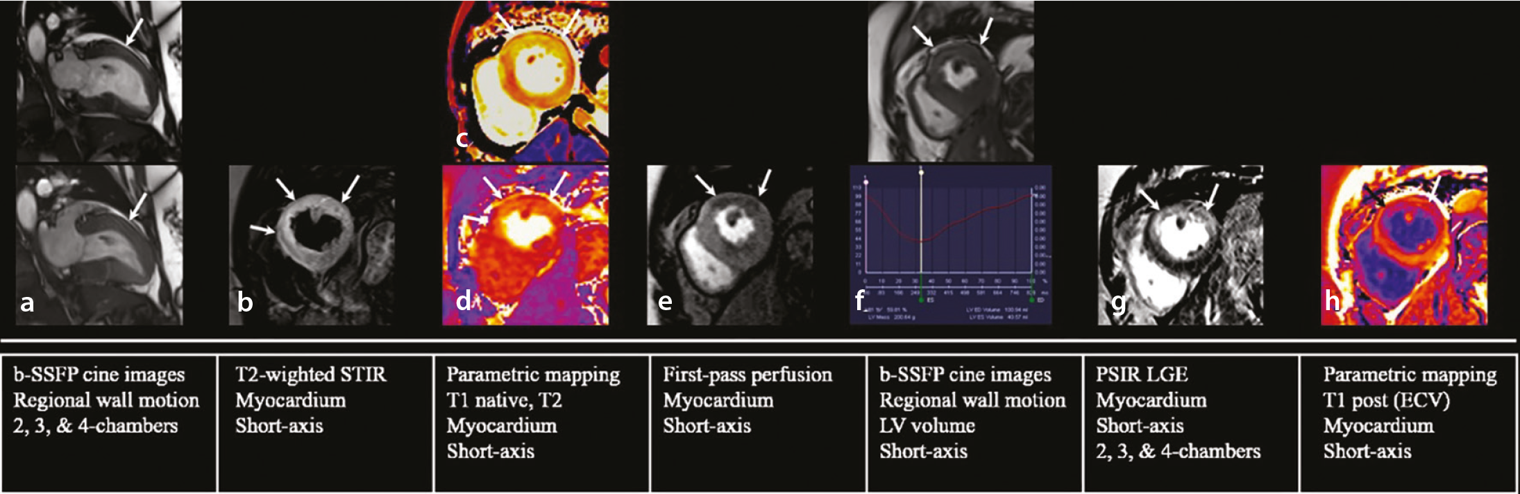
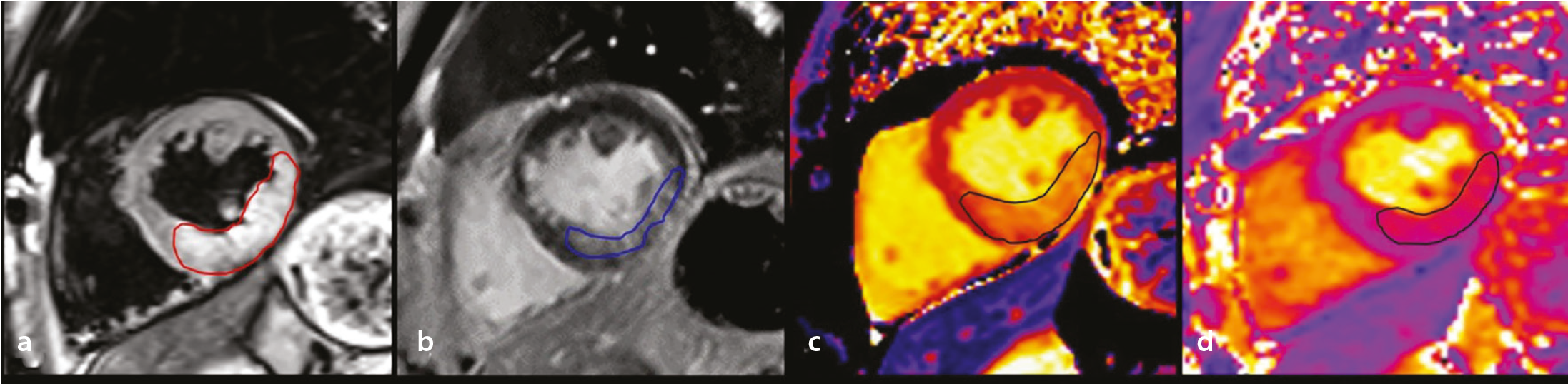
CMR imaging enables a comprehensive evaluation of MI through a multiparametric approach. Standard protocols integrate cine imaging to assess global and regional left ventricular (LV) function; T2-weighted imaging (T2WI) and T2 mapping for myocardial edema and area at risk (AAR); native T1 mapping and extracellular volume (ECV) quantification for diffuse myocardial injury; first-pass perfusion imaging for microvascular integrity; late gadolinium enhancement (LGE) for infarct size, transmural extent, and microvascular obstruction (MVO); and T2* mapping for intramyocardial hemorrhage (IMH). This integrated examination allows for the simultaneous assessment of myocardial injury and salvage and prognostic markers in a single session ().
Quantitative cardiac magnetic resonance imaging biomarkers and prognostic implications
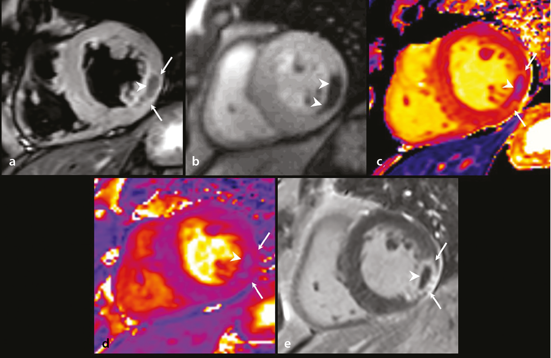
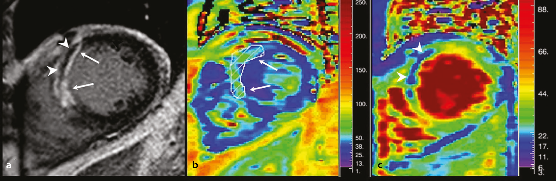
Quantitative CMR imaging biomarkers provide clinically relevant prognostic information beyond a visual assessment. Elevated native T1 values (e.g., > 1,250 ms at 1.5 T) and an increased ECV (> 30%) have been associated with adverse LV remodeling. MVO, visualized as a non-enhancing region within the infarcted myocardium, is a strong predictor of poor outcome, with an MVO burden exceeding approximately 1.4% of the LV mass linked to an increased risk of heart failure and major adverse cardiovascular events (MACE) (). Moreover, T2* values below 20 ms indicate an IMH and are associated with unfavorable remodeling ().,, Texture analysis and radiomics applied to LGE and mapping images have shown promise for predicting LV remodeling and MACE, though these techniques remain investigational.
Myocardial viability, ischemia, and stress cardiac magnetic resonance imaging
LGE-CMR imaging remains the reference standard for the assessment of myocardial viability. Segments with < 25% of LGE involvement demonstrate a high likelihood of functional recovery, whereas those with > 75% involvement rarely recover. Stress CMR imaging, and particularly adenosine stress perfusion imaging, enables a robust functional assessment of myocardial ischemia and may be useful for evaluating residual ischemia and non-culprit lesions after MI, and T2 mapping and T2WI help differentiate acute from chronic MI by depicting infarct-related myocardial edema ().
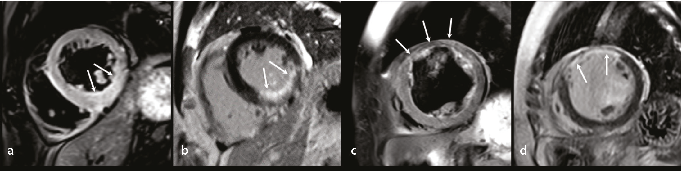
(c, d): Short-axis T2-weighted STIR (c) and LGE (d) images show a normal T2 signal with myocardial thinning (c) and transmural delayed enhancement (d) in the mid-anterior LV segment (arrows), consistent with chronic MI. Although T2-weighted STIR alone cannot reliably distinguish acute from chronic MI, the combination of LGE and T2-weighted STIR enables the identification of infarct-related myocardial edema indicative of acute MI. CMR, cardiac magnetic resonance; MI, myocardial infarction; LV, left ventricular; LGE, late gadolinium enhancement; STIR, short tau inversion recovery.
Area at risk and myocardial salvage
A primary goal in MI management is to limit infarct size and preserve the AAR. In acute MI, T2WI displays myocardial edema that typically extends beyond the infarct core identified on LGE, with the difference representing salvageable myocardium (). Furthermore, T1 and T2 mapping enable quantitative assessment of the AAR and myocardial salvage, thus reducing observer dependency and improving reproducibility. A greater myocardial salvage index has been associated with favorable LV remodeling and functional recovery, highlighting the clinical relevance of mapping-based assessment in post-infarction risk stratification and therapeutic decision making.,
Differentiation between microvascular obstruction, intramyocardial hemorrhage, and myocardial dissecting hematoma
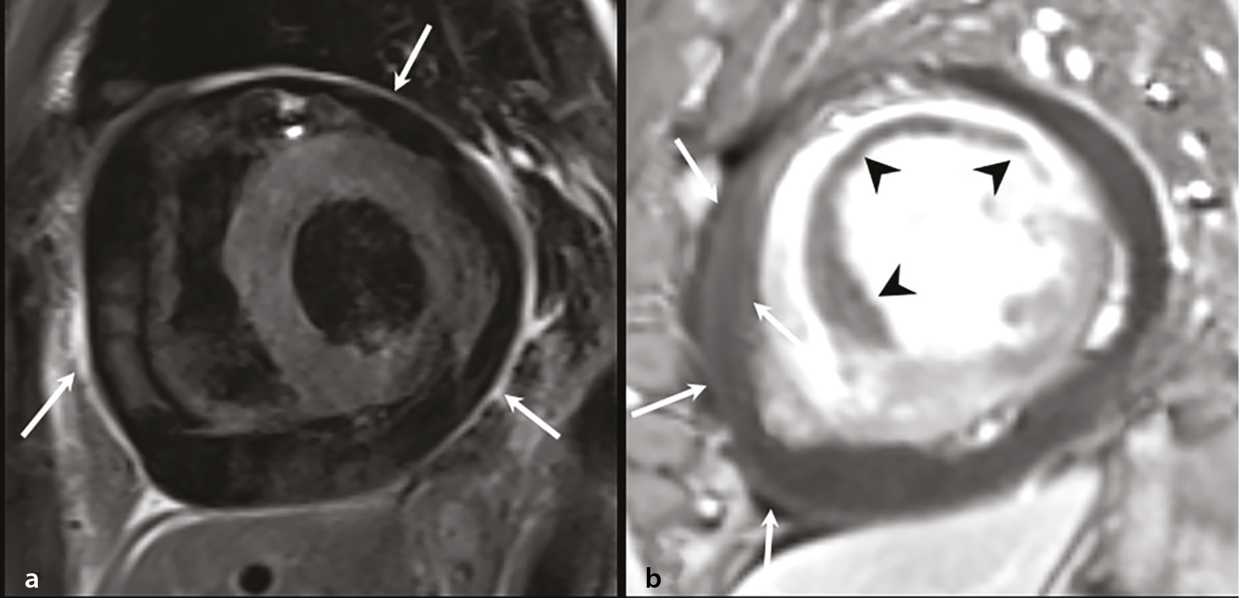
CMR imaging plays a critical role in differentiating between MVO, IMH, and myocardial dissecting hematomas, and accurate distinction between them directly influences their management. MVO appears as a non-enhancing area within the hyperenhanced, infarcted myocardium on LGE and is best delineated on first-pass perfusion imaging. IMH typically co-localizes with MVO and is characterized by shortened T2* values (< 20 ms) resulting from hemosiderin deposition ( and ). By contrast, a myocardial dissecting hematoma presents as an intramyocardial cavity with variable signal intensity, a preserved epicardial contour, and a lack of typical infarct-related enhancement, allowing for its differentiation from reperfusion-related injury and thus guiding appropriate clinical management.
Cardiac computed tomography
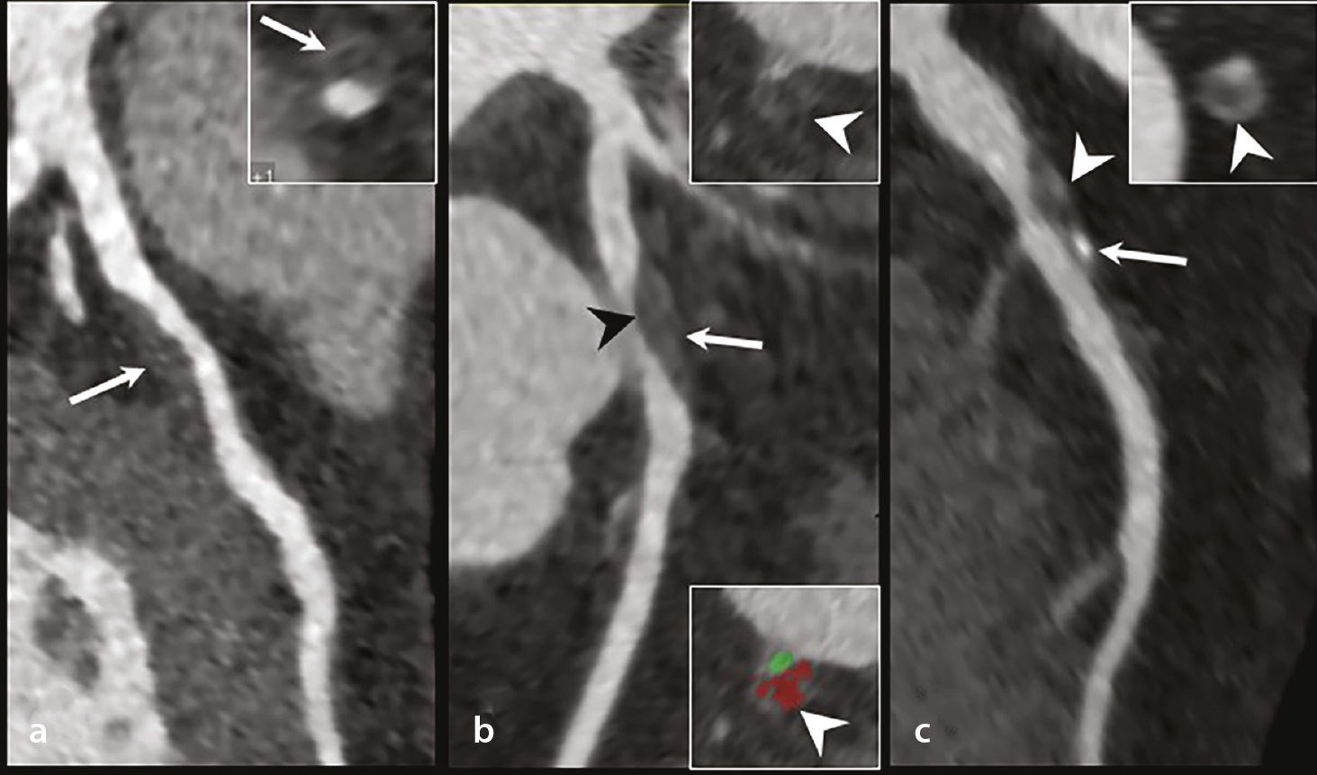
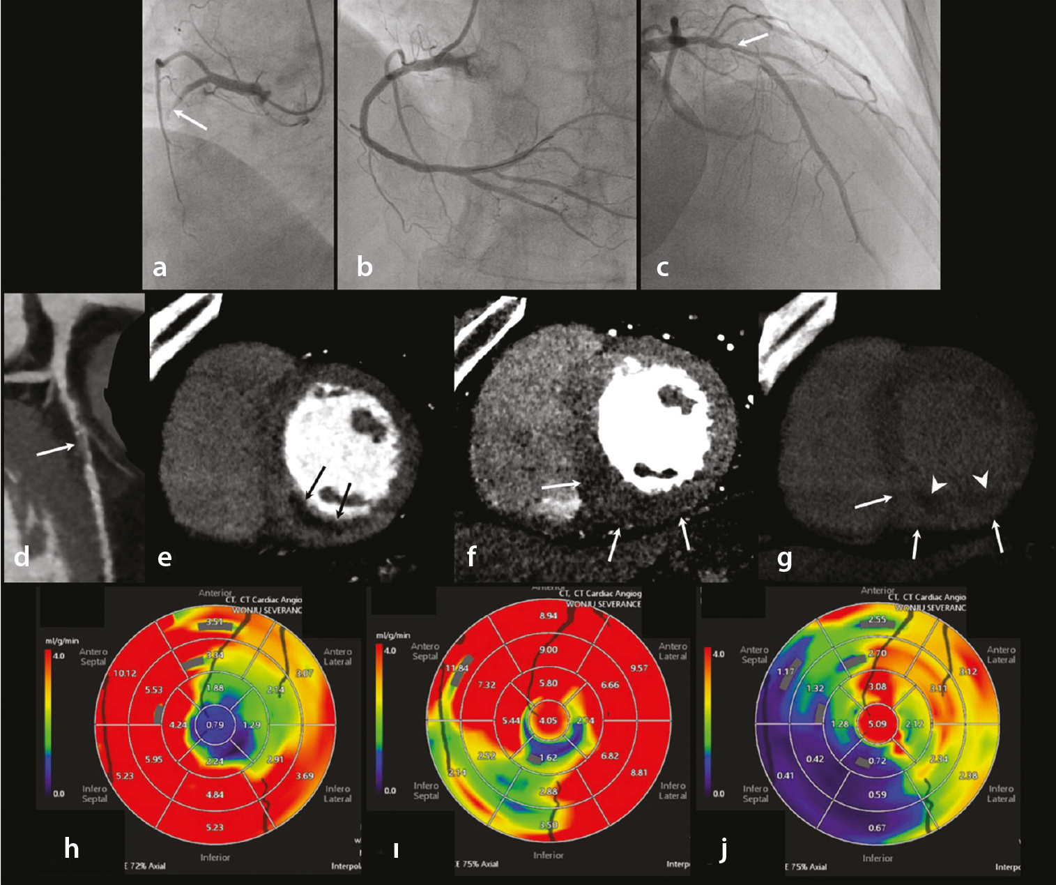
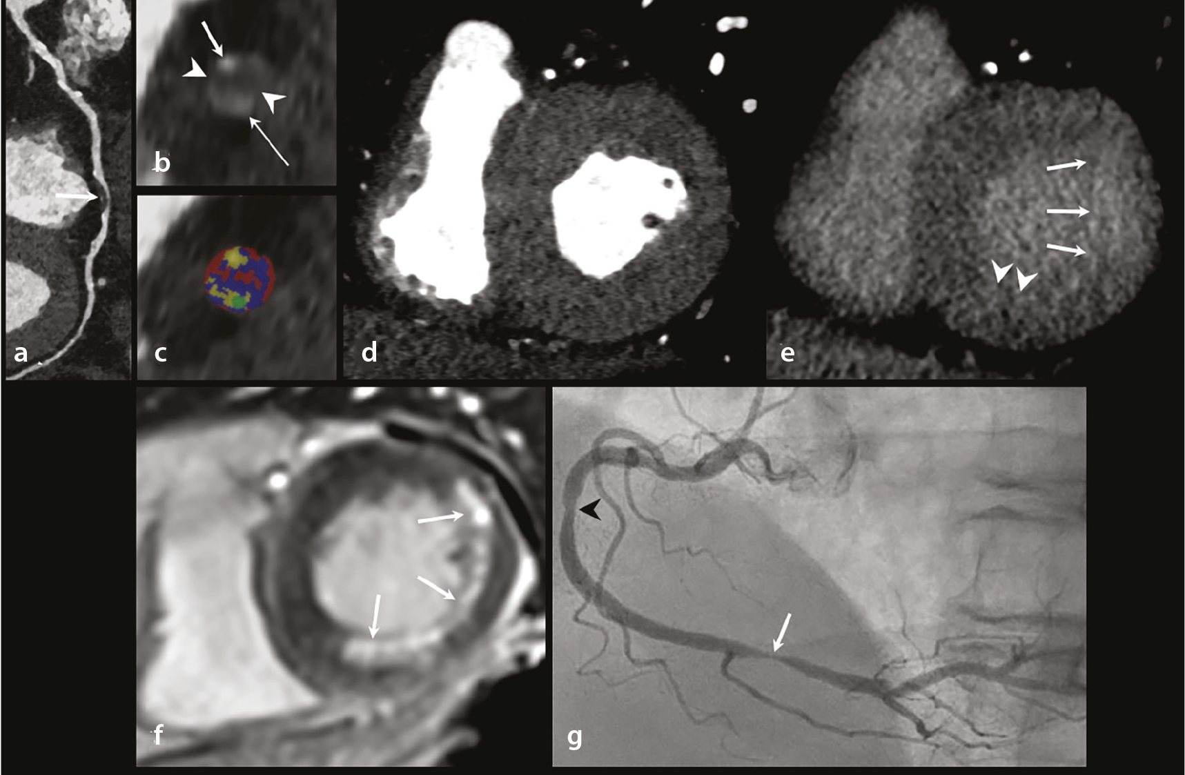
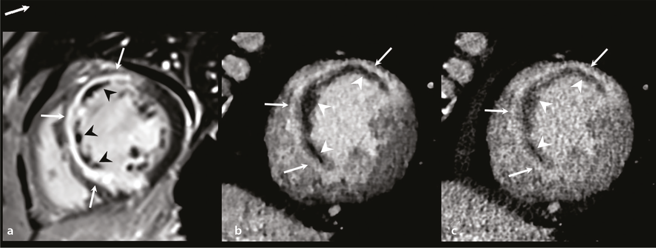
Coronary CT angiography (CCTA) allows for a rapid, non-invasive evaluation of coronary stenosis and plaque morphology, including high-risk features associated with acute coronary syndromes (). When integrated with CCTA, CT-MPI enables combined anatomic and functional assessment and may be useful for evaluating residual ischemia and non-culprit lesions after MI treated with percutaneous coronary intervention. Dynamic stress and rest CT-MPI examinations enable a quantitative assessment of myocardial blood flow and coronary flow reserve, with reduced coronary flow reserve values (typically < 2.0) commonly used as a marker of myocardial ischemia (). MDE-CT visualizes the infarcted myocardium as delayed iodine enhancement and may identify non-enhancing regions consistent with MVO. Compared with LGE-CMR, MDE-CT has inherent limitations, including a lower contrast-to-noise ratio, greater radiation exposure, a lack of standardized acquisition protocols, and limited availability. Accordingly, MDE-CT should be regarded as a complementary and largely investigational technique that is primarily considered when CMR imaging is contraindicated or when a CT scan is already being performed for coronary assessment (). An MDE-CT image is typically acquired 5–10 minutes after contrast administration using low-kV, ECG-triggered delayed scanning with or without a minimal, additional contrast injection. Recent technical advances, including dual-energy iodine mapping and iterative reconstruction, have improved contrast and image quality. In addition, AI-based deep learning denoising enables automated infarct segmentation, enhances the visualization capabilities of MDE, and may facilitate an acceptable image quality at lower radiation doses (). Despite these advances, further external validation and protocol standardization are required before broader clinical adoption can proceed.
Imaging of post-myocardial infarction complications
A variety of mechanical and thromboembolic complications may occur after MI, particularly with delayed or inadequate reperfusion, and are associated with hemodynamic instability and adverse outcomes.,
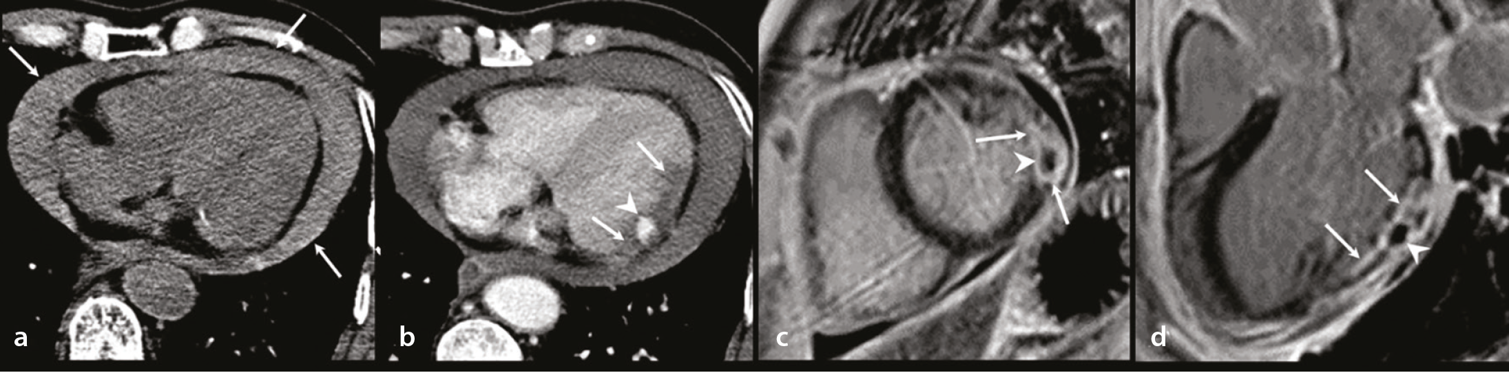
Free wall rupture is a catastrophic complication of STEMI. CCT enables rapid assessment, displaying hemopericardium, pericardial effusion, and rupture tracts, whereas LGE-CMR confirms myocardial discontinuity, irregular margins, and associated thrombi, facilitating prompt diagnosis and management ().
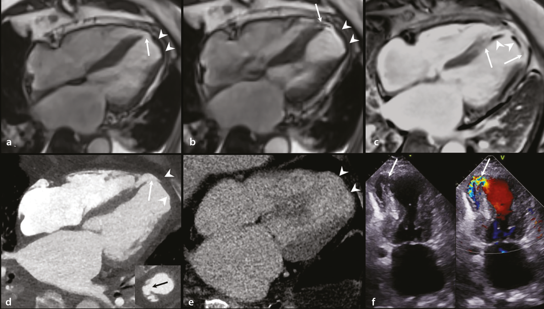
Ventricular septal rupture is an uncommon but life-threatening complication of MI. CCT provides the rapid, non-invasive detection of septal defects, whereas CMR imaging offers a detailed evaluation of defect size and location and surrounding myocardial integrity, which is essential for surgical planning and prognostic assessment ().
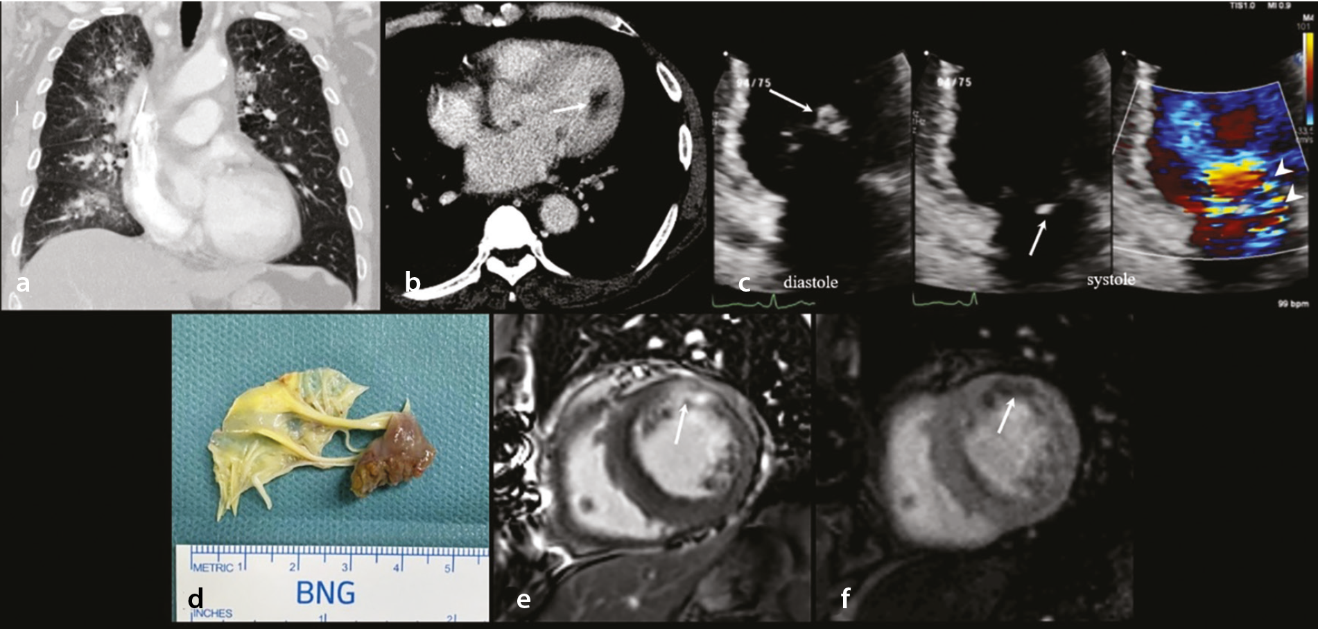
Papillary muscle rupture results in acute, severe mitral regurgitation. Echocardiography remains the first-line modality for hemodynamic assessment, whereas CCT and CMR imaging allow for the visualization of papillary muscle integrity and global LV function. In chronic mitral regurgitation, CMR imaging enables an accurate quantification of regurgitant volume and an assessment of myocardial fibrosis using LGE and T1 mapping ().
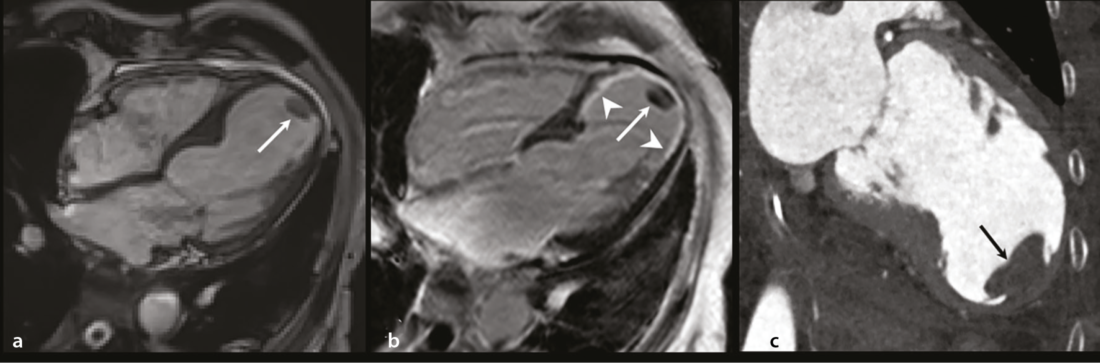
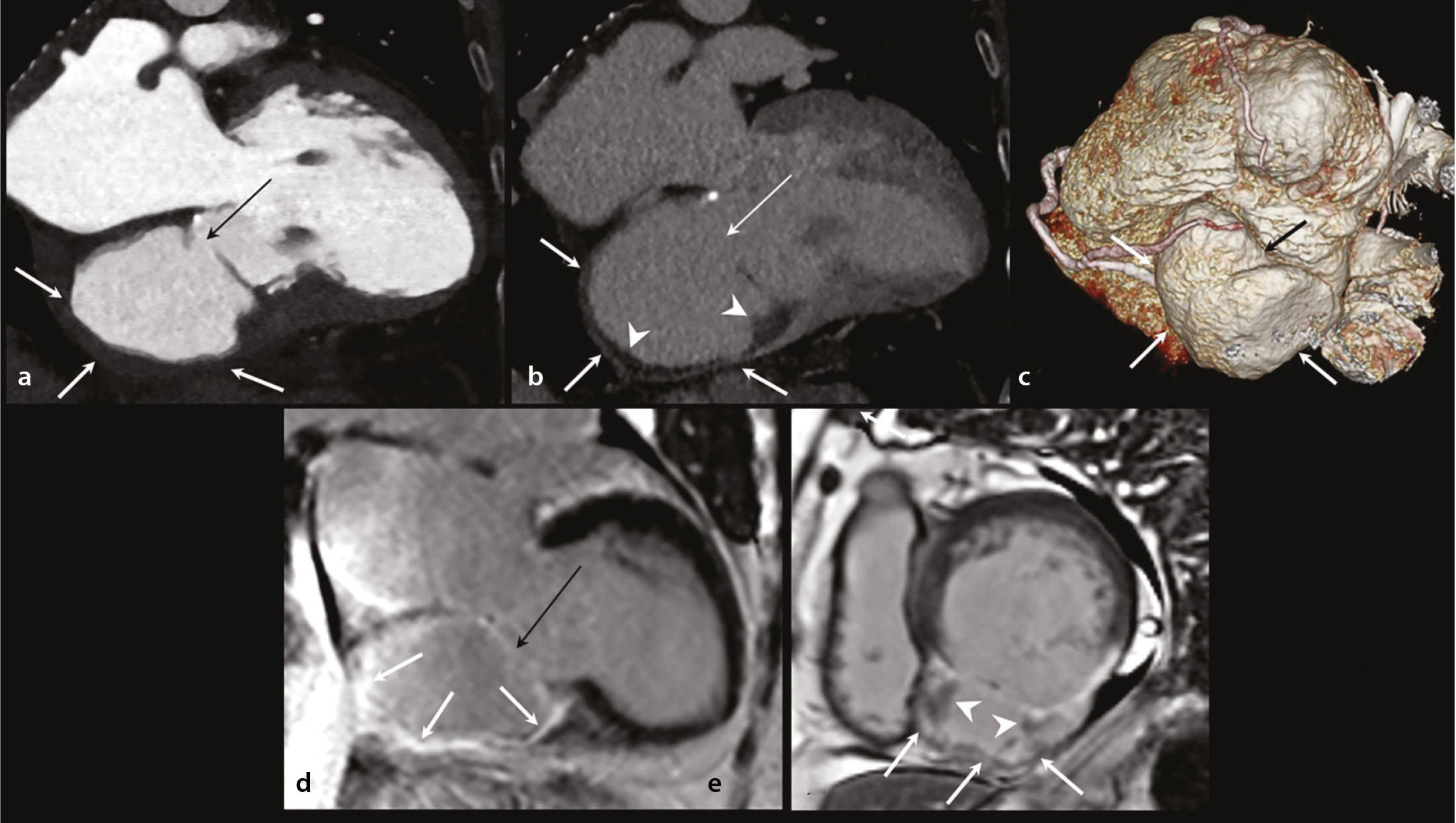
Differentiation between a true aneurysm and a pseudoaneurysm relies heavily on imaging. CCT and CMR imaging assess wall continuity, neck morphology, thrombus formation, and pericardial involvement, which are critical for guiding management decisions ( and ).
LV thrombi are a common post-MI complication. On LGE-CMR, a thrombus appears as a non-enhanced filling defect, with long inversion time imaging improving thrombus conspicuity by suppressing myocardial and blood pool signals. Cine imaging allows for the assessment of thrombus mobility, which aids risk stratification and anticoagulation planning ().
Pericardial complications, including pericarditis and effusion, are readily evaluated with CCT for anatomic assessment. CMR imaging enables characterization of pericardial inflammation and enhancement.
Heart failure represents a major long-term consequence of MI. CMR imaging enables a comprehensive evaluation of ventricular function, valvular mechanics, and myocardial tissue characteristics to support longitudinal assessment and management.
Clinical integration and modality selection
Rather than competing modalities, CCT, CMR imaging, and echocardiography provide synergistic information., , , Modality selection should be guided by the clinical question at hand, patient stability, contraindications, and local expertise. A practical clinical algorithm summarizing modality choice is provided in .
Table 1. Algorithm for clinical imaging modality selection following myocardial infarction
Clinical scenario | Preferred modality | Rationale |
|---|---|---|
Hemodynamically unstable acute MI | Echocardiography | Bedside, real-time assessment of ventricular function and mechanical complications. |
Post-reperfusion viability and area at risk | CMR imaging | Gold standard for assessing infarct size, transmurality, area at risk, and MVO. |
Non-culprit lesion significance/residual ischemia | Stress CMR or cardiac CT (CCTA + CT-derived MPI/CT-FFR) | Functional assessment of ischemia with an integrated anatomic–functional assessment. |
Suspected LV thrombus | CMR (long-TI LGE) imaging | Highest diagnostic accuracy for thrombus detection and characterization. |
MRI contraindicated | Cardiac CT (MDE-CT) | Complementary alternative for myocardial viability assessment. |
Mechanical complications | CT + echocardiography + CMR | Comprehensive evaluation of anatomy, hemodynamics, and myocardial integrity. |
Remodeling/prognosis | CMR (T1 mapping, ECV, MVO) | Quantitative biomarkers for risk stratification and outcome prediction. |
MI, myocardial infarction; CMR, cardiac magnetic resonance; MVO, microvascular obstruction; CT, computed tomography; CCTA, coronary CT angiography; MPI, myocardial perfusion imaging; FFR, fractional flow reserve; LV, left ventricular; TI, inversion time; LGE, late gadolinium enhancement; MRI, magnetic resonance imaging; MDE, myocardial delayed enhancement; ECV, extracellular volume.
Ethics and consent
This pictorial essay was conducted in accordance with institutional ethics standards. Formal institutional review board approval and informed consent were waived owing to the essay’s retrospective use of anonymized data.
This pictorial essay does not provide pooled diagnostic accuracy metrics, and quantitative thresholds may vary by scanner, field strength, and post-processing procedures. Advanced CT techniques, such as MDE-CT and CT-MPI, and emerging AI-based and radiomics approaches are not yet widely standardized—a circumstance that could potentially limit the generalizability of this essay’s conclusions. Therefore, this article focuses on established and clinically applicable imaging findings.
CCT and CMR imaging provide complementary information in the evaluation of MI beyond the acute phase. CMR imaging enables detailed tissue characterization, viability assessment, and evaluation of post-infarction complications. CCT allows for rapid coronary assessment and functional evaluation and plays an important role in assessing select post-infarction complications. Integrated with echocardiography and clinical context, multimodality imaging allows for the characterization of infarct-related injury and residual ischemia. A scenario-based imaging approach supports informed clinical decision-making in patients with MI.
Footnotes
Conflict of interest disclosure
The authors declared no conflicts of interest.
Funding
This work was supported by the Natural Science Foundation of Shandong Province.
References
Salari N, Morddarvanjoghi F, Abdolmaleki A, et al. The global prevalence of myocardial infarction: a systematic review and meta-analysis. BMC Cardiovasc Disord. 2023;23(1):206.
Rajiah P, Desai MY, Kwon D, Flamm SD. MR imaging of myocardial infarction. RadioGraphics. 2013;33(5):1383-1412.
Peper J, Suchá D, Swaans M, Leiner T. Functional cardiac CT-going beyond anatomical evaluation of coronary artery disease with cine CT, CT-FFR, CT perfusion and machine learning. Br J Radiol. 2020;93(1113):20200349.
Williams MC, Weir-McCall JR, Baldassarre LA, et al. Artificial intelligence and machine learning for cardiovascular computed tomography (CCT): A White Paper of the Society of Cardiovascular Computed Tomography (SCCT). J Cardiovasc Comput Tomogr. 2024;18(6):519-532.
Thygesen K, Alpert JS, Jaffe AS, et al. Fourth universal definition of myocardial infarction (2018). Circulation. 2018;138(20):e618-e651.
Kloner RA. Stunned and hibernating myocardium: where are we nearly 4 decades later? J Am Heart Assoc. 2020;9(3):e015502.
Bulluck H, Dharmakumar R, Arai AE, Berry C, Hausenloy DJ. Cardiovascular magnetic resonance in acuteST-segment-elevation myocardial infarction: recent advances, controversies, and future directions. Circulation. 2018;137(18):1949-1964.
Liu D, Borlotti A, Viliani D, et al. CMR native T1 mapping allows differentiation of reversible versus irreversible myocardial damage in ST-segment-elevation myocardial infarction: an OxAMI Study (Oxford Acute Myocardial Infarction). Circ Cardiovasc Imaging. 2017;10(8):e005986.
Chen Y, Ren D, Guan X, et al. Quantification of myocardial hemorrhage using T2* cardiovascular magnetic resonance at 1.5T with ex-vivo validation. J Cardiovasc Magn Reson. 2021;23(1):104.
Bulluck H, Carberry J, Carrick D, et al. A noncontrast CMR risk score for long-term risk stratification in reperfused ST-segment elevation myocardial infarction. JACC Cardiovasc Imaging. 2022;15(3):431-443.
Ma Q, Ma Y, Wang X, et al. A radiomic nomogram for prediction of major adverse cardiac events in ST-segment elevation myocardial infarction. Eur Radiol. 2021;31(2):1140-1150.
Garcia MJ, Kwong RY, Scherrer-Crosbie M, et al. State of the art: imaging for myocardial viability: a scientific statement from the American Heart Association. Circ Cardiovasc Imaging. 2020;13(7):e000053.
Nagel E, Greenwood JP, McCann GP, et al. Magnetic resonance perfusion or fractional flow reserve in coronary disease. N Engl J Med. 2019;380(25):2418-2428.
Alyousef T, Malhotra S, Iskander F, et al. Left ventricular intramyocardial dissecting hematoma: a multimodality imaging diagnostic approach. Circ Cardiovasc Imaging. 2021;14(7):e012410.
Pontone G, Baggiano A, Andreini D, et al. Stress computed tomography perfusion versus fractional flow reserve CT derived in suspected coronary artery disease: the PERFECTION Study. JACC Cardiovasc Imaging. 2019;12(8 Pt 1):1487-1497.
Dewey M, Siebes M, Kachelrieß M, et al. Clinical quantitative cardiac imaging for the assessment of myocardial ischaemia. Nat Rev Cardiol. 2020;17(7):427-450.
Rodriguez-Granillo GA. Delayed enhancement cardiac computed tomography for the assessment of myocardial infarction: from bench to bedside. Cardiovasc Diagn Ther. 2017;7(2):159-170.
Nishii T, Kobayashi T, Tanaka H, et al. Deep Learning-based post hoc CT denoising for myocardial delayed enhancement. Radiology. 2022;305(1):82-91.
Stephens NR, Restrepo CS, Saboo SS, Baxi AJ. Overview of complications of acute and chronic myocardial infarctions: revisiting pathogenesis and cross-sectional imaging. Postgrad Med J. 2019;95(1126):439-450.
Damluji AA, van Diepen S, Katz JN, et al. Mechanical complications of acute myocardial infarction: a scientific statement from the American Heart Association. Circulation. 2021;144(2):e16-e35.