
Dear Editor,
We would like to thank Dr. Kaba for his kind and insightful comments on our article “Single-center retrospective comparative study of 40–120 μm versus 100–300 μm trisacryl gelatin microspheres as the main embolic agents for prostatic artery embolization”, which was recently published in Diagnostic and Interventional Radiology.
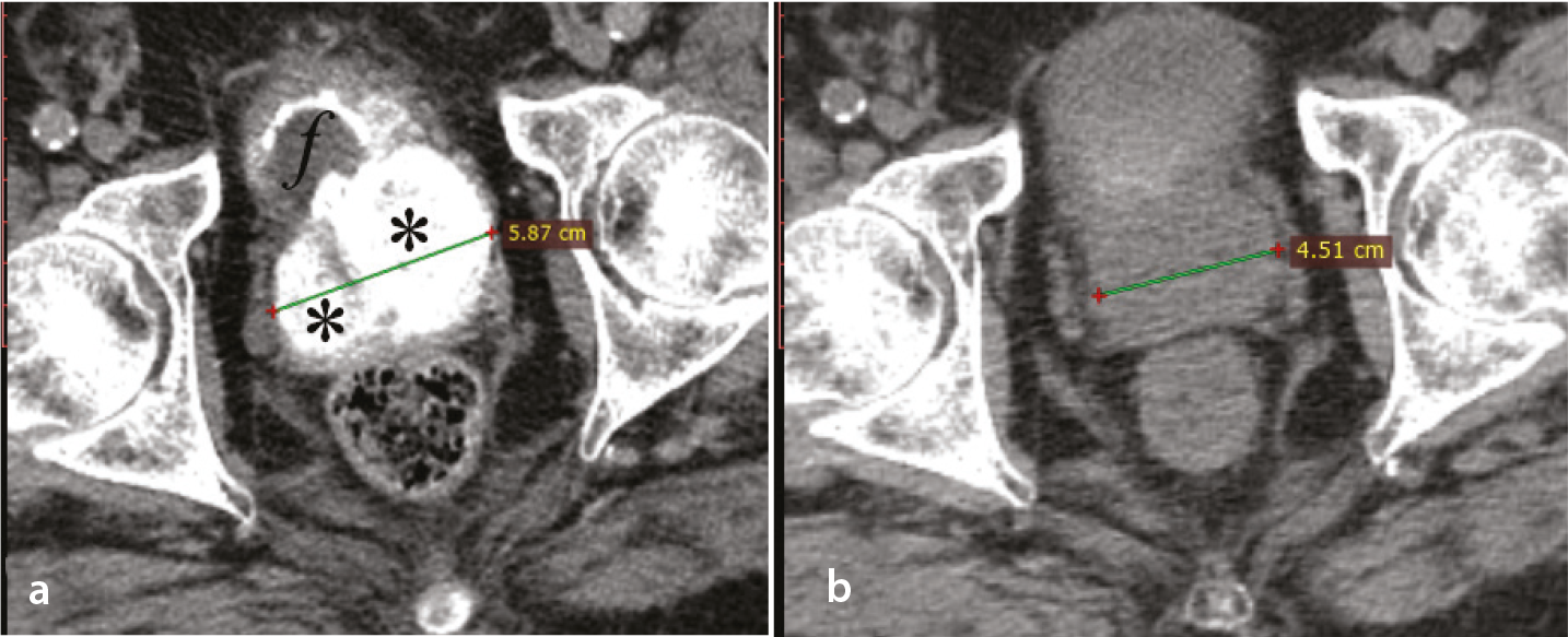
We fully agree with Dr. Kaba that, during prostatic artery embolization (PAE) with small (Embosphere 40–120 μm or equivalent) microspheres, a delicate balance must be maintained between efficacy (strong local ischemic effect) and safety (ischemic complications). As noted in the letter, the operator has to decide between maximal prostatic ischemia (potentially associated with severe intraprocedural pain and harmful, non-target embolization) and sufficient ischemia (with lower respective risks). But how can we measure and define maximal and sufficient prostatic ischemia? Regarding maximal ischemia, our experience with utilization of small microspheres and contrast-enhanced ultrasound (CEUS) for early post-PAE measurement of the percentage of prostatic ischemia (pPI), shows that up to 80% of the total prostate volume can be devascularized and appear as non-enhancing areas on CEUS or as extensive hyperdensity of prostate parenchyma on non-enhanced computed tomography (CT) (). Such extensive ischemia is usually associated with significant prostate shrinkage (up to 65%, 3–6 months post-PAE) and with durable clinical improvement. Unfortunately, there is scarce evidence regarding sufficient prostatic ischemia. In patients with an indwelling bladder catheter (IBC), it has been demonstrated that successful removal of the catheter could be achieved only in patients with pPI > 10% (measured by CEUS 1 day post-PAE).
Interestingly, a similar conclusion was reached by a magnetic resonance (MR) study of prostatic infarction post-PAE in patients with an IBC. Another MR study reported better 12-month clinical outcomes in patients with infarction of > 5%; however, infarction was assessed 3 months post-PAE. Intraprocedural evaluation of infarction with CEUS performed in the angio-suite shortly after injection of the embolic agent is an advantage of CEUS over MR and probably deserves further investigation and clinical correlation. Such an evaluation could be used to guide on-site treatment decisions and help the operator maintain the aforementioned delicate balance.
We also agree that cone-beam CT should be systematically applied during PAE with small microspheres in order to reveal communication of the prostatic artery with the middle rectal artery and other small (but potentially important) neighboring splanchnic pelvic branches. As additional measures to reduce the prevalence and severity of intraprocedural and postprocedural complications, we propose: 1) strong intravenous analgesia (e.g., 50 mcg fentanyl + 1 mg midazolam administered 10 minutes before arterial puncture) and intraarterial administration of lidocaine hydrochloride through the microcatheter just before injection of the microspheres; and 2) use of a balloon-occlusion microcatheter when there is a concern for reflux of embolic agent into the superior vesical, internal pudendal, or middle rectal artery.
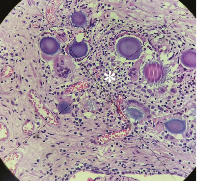
Finally, we would like to provide some histologic evidence for the distal penetration of 40–120 μm microspheres () and to underline the need for additional relevant studies, with longer follow-up, that could reveal a clinical benefit for this type of microsphere.
Footnotes
Conflict of interest disclosure
The authors declared no conflicts of interest.
References
Kaba E. Letter to the Editor: small microspheres for prostatic artery embolization: balancing efficacy and safety. Diagn Interv Radiol. 2026.
Moschouris H, Şentürk Ç, Stamatiou K, Mouroukis A, Stylianou C, Kontothanasis D. Single-center retrospective comparative study of 40-120 μm versus 100-300 μm trisacryl gelatin microspheres as the main embolic agents for prostatic artery embolization. Diagn Interv Radiol. 2026;32(5):700-708.
Moschouris H, Stamatiou K, Malagari K, et al. The value of contrast-enhanced ultrasonography in detection of prostatic infarction after prostatic artery embolization for the treatment of symptomatic benign prostatic hyperplasia. Diagn Interv Radiol. 2019;25(2):134-143.
Kisilevzky N, Faintuch S. MRI assessment of prostatic ischaemia: best predictor of clinical success after prostatic artery embolisation for benign prostatic hyperplasia. Clin Radiol. 2016;71(9):876-882.
von Stempel C, Tzelves L, Khangura S, et al. Three-month magnetic resonance imaging post-prostate artery embolization predicts 1-year clinical outcomes. Cardiovasc Intervent Radiol. 2026;49(2):370-379.
Moschouris H, Stamatiou K. Intentionally unilateral prostatic artery embolization: patient selection, technique and potential benefits. World J Radiol. 2024;16(9):380-388.